Copyright
©The Author(s) 2020.
World J Gastroenterol. Jul 21, 2020; 26(27): 3899-3916
Published online Jul 21, 2020. doi: 10.3748/wjg.v26.i27.3899
Published online Jul 21, 2020. doi: 10.3748/wjg.v26.i27.3899
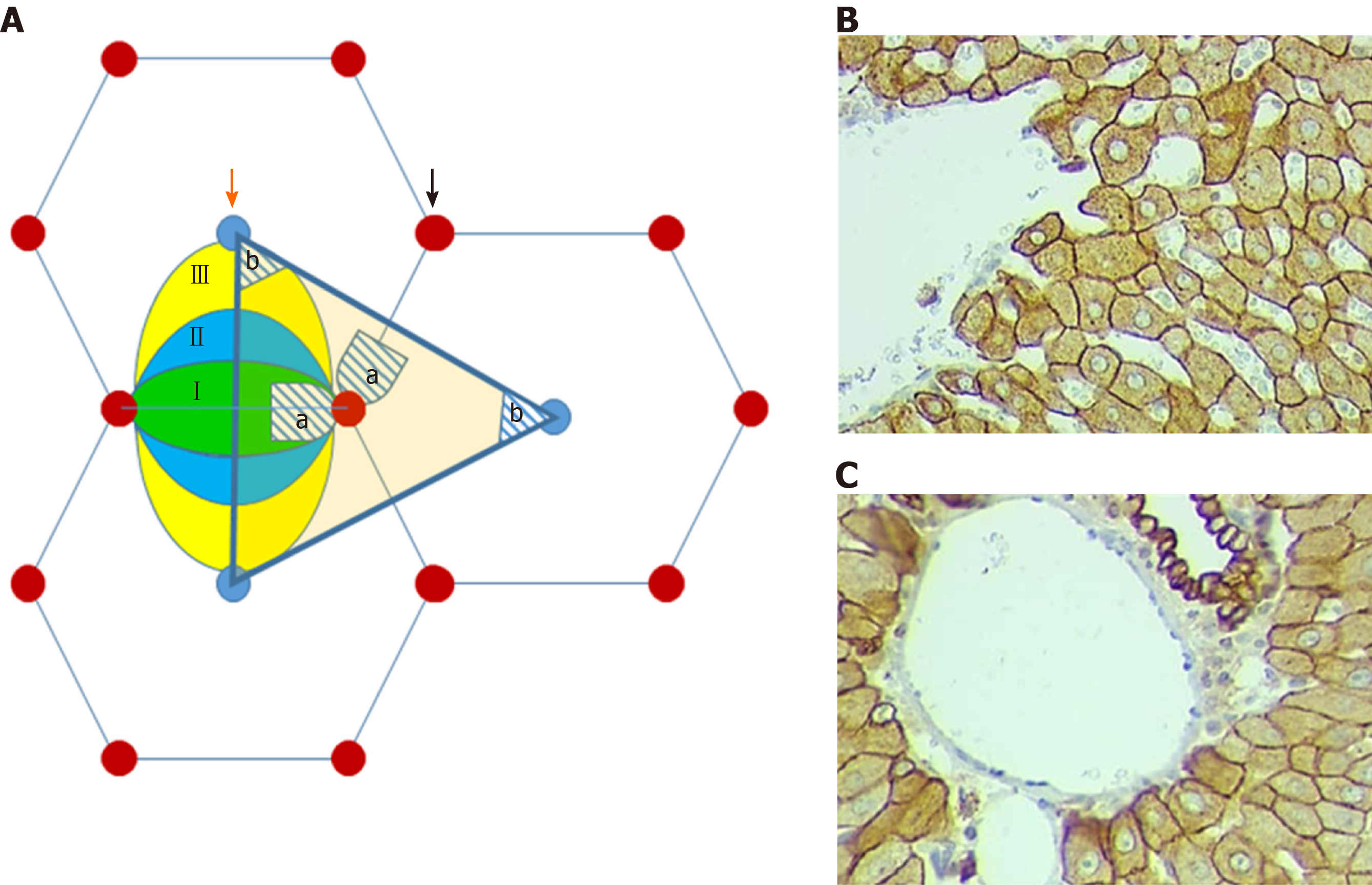
Figure 1 Schematic and histological figures of liver lobule.
A: Schematic illustration of mutual compatibility of “classical” and “portal” lobules with hepatic acini. I, II, III – the zones of the acinus. The hatched periportal area “a” – localized in the 1st zone of the acinus. The hatched pericentral area “b” – localized in the 3rd zone of the acinus. Orange arrow – central vein; black arrow – portal triad. B: Pericentral hepatocytes (CK8) ObX40; OcX20; C: Periportal hepatocytes (CK8). ObX40; OcX20.
- Citation: Tsomaia K, Patarashvili L, Karumidze N, Bebiashvili I, Azmaipharashvili E, Modebadze I, Dzidziguri D, Sareli M, Gusev S, Kordzaia D. Liver structural transformation after partial hepatectomy and repeated partial hepatectomy in rats: A renewed view on liver regeneration. World J Gastroenterol 2020; 26(27): 3899-3916
- URL: https://www.wjgnet.com/1007-9327/full/v26/i27/3899.htm
- DOI: https://dx.doi.org/10.3748/wjg.v26.i27.3899


