Copyright
©The Author(s) 2020.
World J Gastroenterol. Jul 7, 2020; 26(25): 3660-3672
Published online Jul 7, 2020. doi: 10.3748/wjg.v26.i25.3660
Published online Jul 7, 2020. doi: 10.3748/wjg.v26.i25.3660
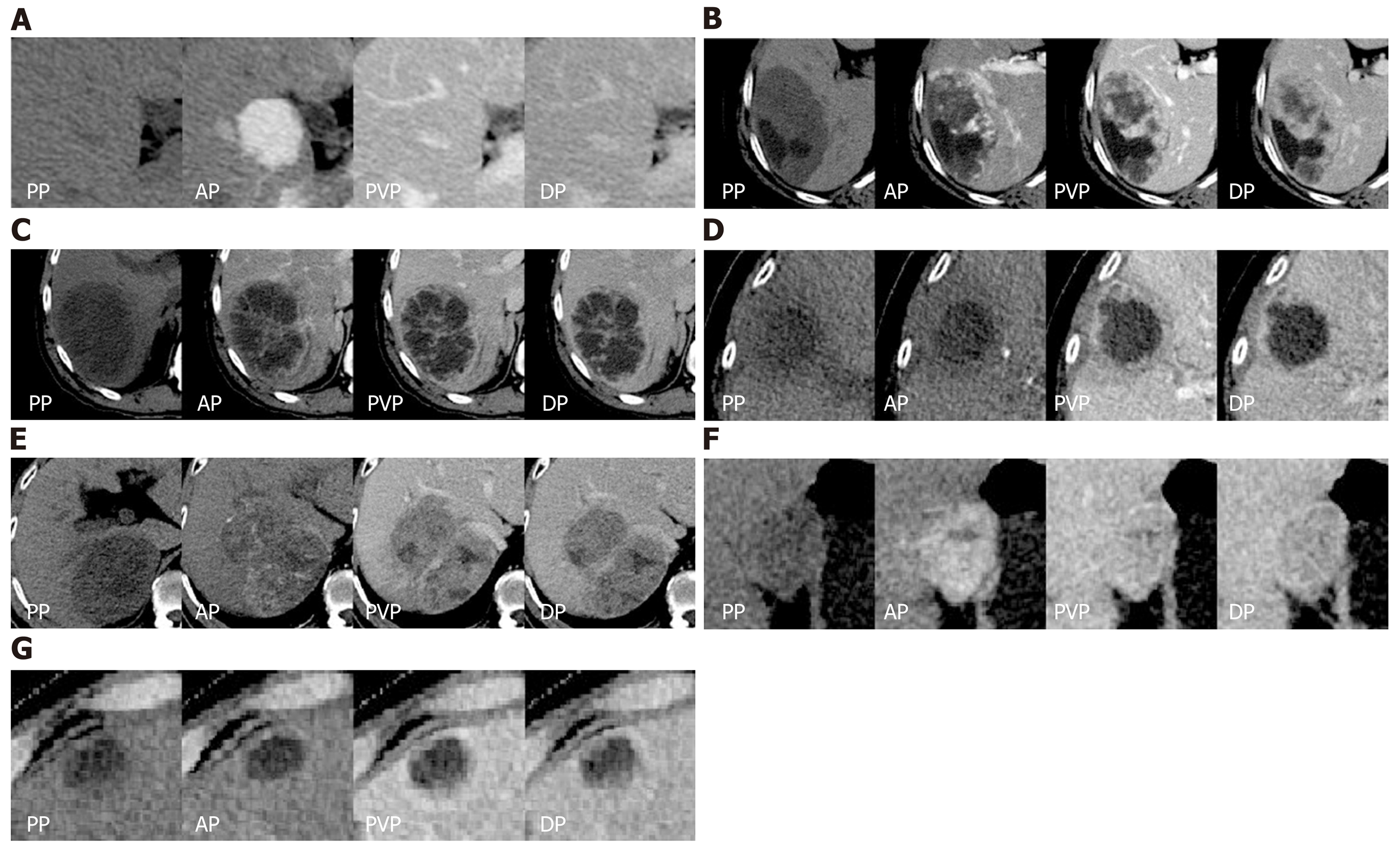
Figure 3 The representative correctly classified and misclassified categories.
For each patient, axial four-phase (PP, AP, PVP, DP) computed tomography images were obtained and focal liver lesions were diagnosed by histopathologic evaluation after biopsy or surgery. A: A 33-year-old man with focal nodular hyperplasia was correctly classified as category C; B: A 54-year-old woman with hemangioma was misclassified as category D; C: A 52-year-old man with hepatic abscess was correctly classified as category D; D: An 82-year-old woman with hepatic abscess was misclassified as category B; E: A 55-year-old man with HCC was correctly classified as category A; F: A 38-year-old woman with HCC was misclassified as category C; G: A 75-year-old man with liver metastases derived from colorectal cancer was correctly classified as category B. And there was no misclassification for the metastasis group. AP: Arterial phase; DP: Delayed phase; PP: Precontrast phase; PVP: Portal venous phase.
- Citation: Cao SE, Zhang LQ, Kuang SC, Shi WQ, Hu B, Xie SD, Chen YN, Liu H, Chen SM, Jiang T, Ye M, Zhang HX, Wang J. Multiphase convolutional dense network for the classification of focal liver lesions on dynamic contrast-enhanced computed tomography. World J Gastroenterol 2020; 26(25): 3660-3672
- URL: https://www.wjgnet.com/1007-9327/full/v26/i25/3660.htm
- DOI: https://dx.doi.org/10.3748/wjg.v26.i25.3660


