Copyright
©The Author(s) 2020.
World J Gastroenterol. Jul 7, 2020; 26(25): 3542-3561
Published online Jul 7, 2020. doi: 10.3748/wjg.v26.i25.3542
Published online Jul 7, 2020. doi: 10.3748/wjg.v26.i25.3542
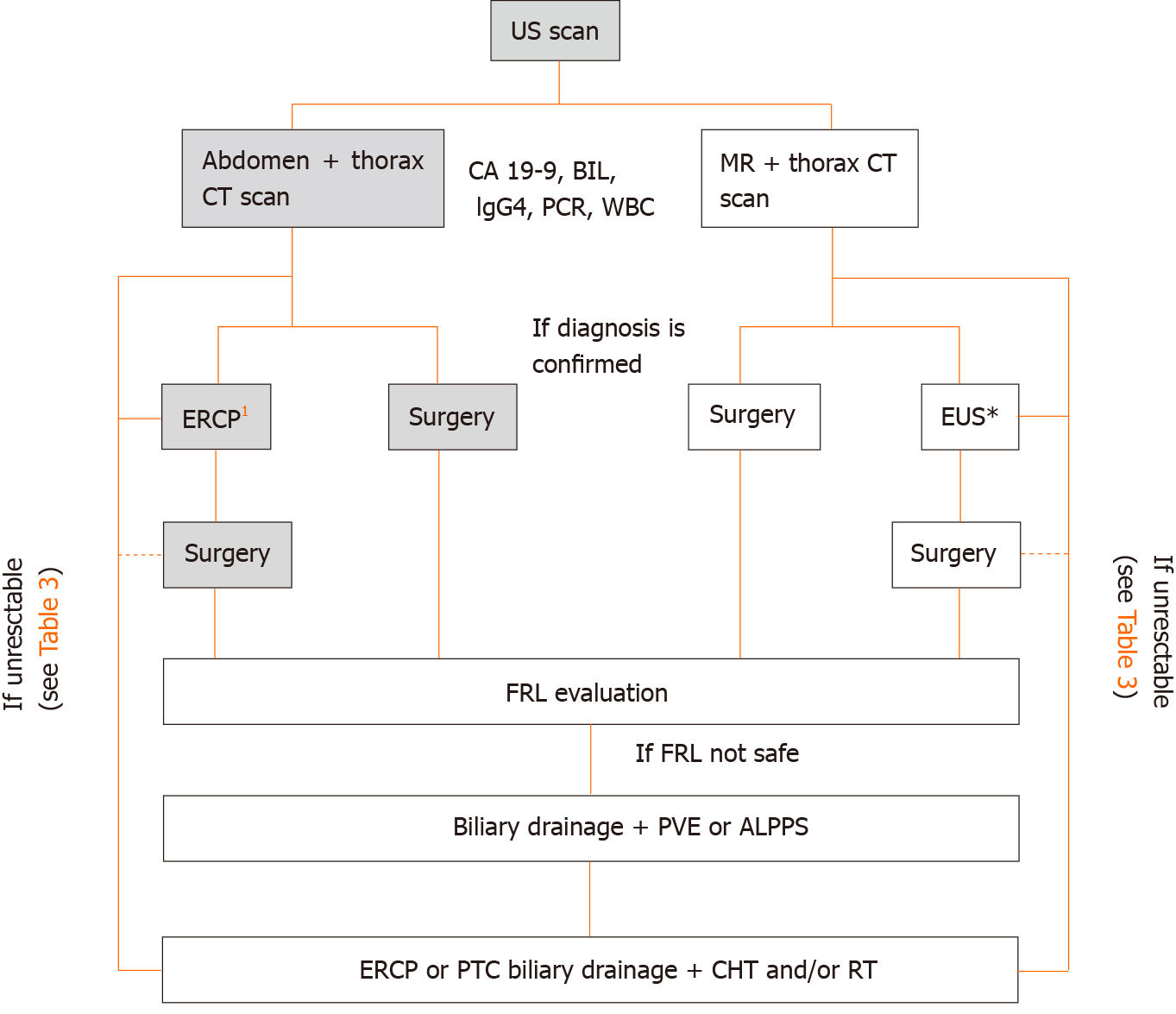
Figure 2 Diagnostic and therapeutic work-flow for perihilar cholangiocarncioma.
1If cytological confirmation is needed (negative carbohydrate antigen 19-9, positive immunoglobulin G4, and confounding diagnosis at imaging); interrupted line, consider neo-adjuvant therapies. US: Ultrasound; CT: Computed tomography; MR: Magnetic resonance; CA 19-9: Carbohydrate antigen 19-9; BIL: Bilirubin; IgG4: Immunoglobulin G4; ERCP: Endoscopic-retrograde-pancreatoduodenoscopy; EUS: Endoscopic ultrasound; FRL: Future remnant liver; PVE: Portal vein embolization; ALPPS: Associated liver partition to portal vein ligation for staged hepatectomy; PTC: Percutaneous transhepatic colangiography; CHT: Chemotherapy; RT: Radiotherapy.
- Citation: Dondossola D, Ghidini M, Grossi F, Rossi G, Foschi D. Practical review for diagnosis and clinical management of perihilar cholangiocarcinoma. World J Gastroenterol 2020; 26(25): 3542-3561
- URL: https://www.wjgnet.com/1007-9327/full/v26/i25/3542.htm
- DOI: https://dx.doi.org/10.3748/wjg.v26.i25.3542


