Copyright
©The Author(s) 2020.
World J Gastroenterol. Jun 7, 2020; 26(21): 2792-2809
Published online Jun 7, 2020. doi: 10.3748/wjg.v26.i21.2792
Published online Jun 7, 2020. doi: 10.3748/wjg.v26.i21.2792
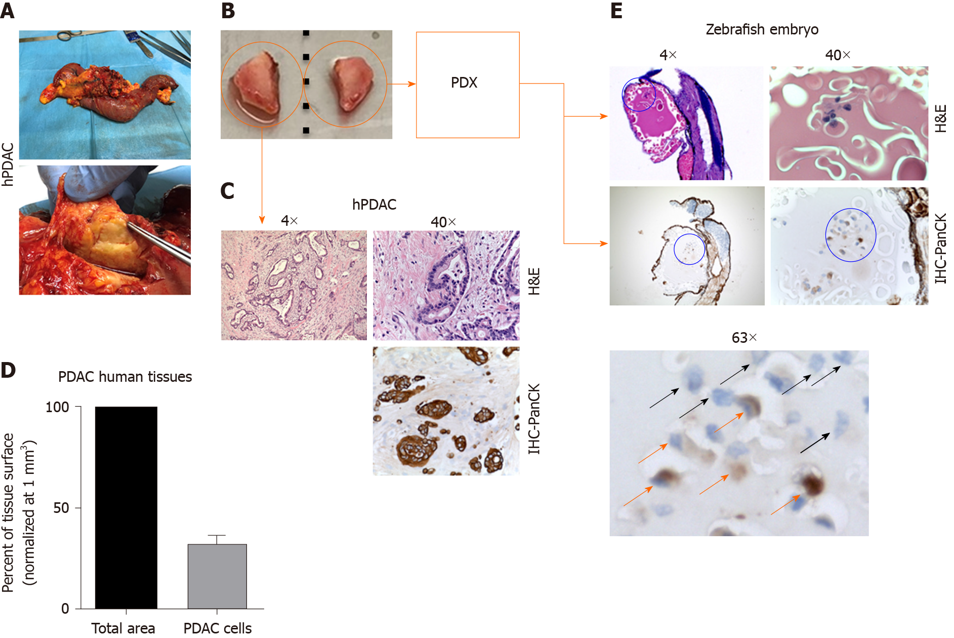
Figure 2 Tumor tissue was successfully engrafted in healthy zebrafish embryos in all cases.
A: Surgical specimen was obtained from patient with diagnosed pancreatic ductal adenocarcinoma (PDAC); B: Two fragments of the tumor were taken; C and D: One of them was used for histological evaluation (C) revealing that the percentage of epithelial cells (mean PDAC counterpart) out of the total surface area is 31.8% ± 4.9% (n = 3) (D); E: The second fragment of the tumor was used for xenotransplantation into the yolk of zebrafish embryos 2 dpf. After 2 d post xenotransplantation, the zebrafish patient-derived xenografts (zPDX) are Formalin-Fixed Paraffin Embedded. We performed the hematoxylin and eosin staining and immunohistochemistry using anti-Human Pan-Cytokeratin antibody on zPDX sections, highlighting the presence of epithelial PDAC cells (orange arrows) and the immune-negative counterpart (black arrows) that might be associated with the microenvironment side of PDAC human tissue. PDAC: Pancreatic ductal adenocarcinoma; PDX: Patient-derived xenografts; PanCK: Pan-Cytokeratin antibody.
- Citation: Di Franco G, Usai A, Funel N, Palmeri M, Montesanti IER, Bianchini M, Gianardi D, Furbetta N, Guadagni S, Vasile E, Falcone A, Pollina LE, Raffa V, Morelli L. Use of zebrafish embryos as avatar of patients with pancreatic cancer: A new xenotransplantation model towards personalized medicine. World J Gastroenterol 2020; 26(21): 2792-2809
- URL: https://www.wjgnet.com/1007-9327/full/v26/i21/2792.htm
- DOI: https://dx.doi.org/10.3748/wjg.v26.i21.2792


