Copyright
©The Author(s) 2020.
World J Gastroenterol. May 28, 2020; 26(20): 2618-2631
Published online May 28, 2020. doi: 10.3748/wjg.v26.i20.2618
Published online May 28, 2020. doi: 10.3748/wjg.v26.i20.2618
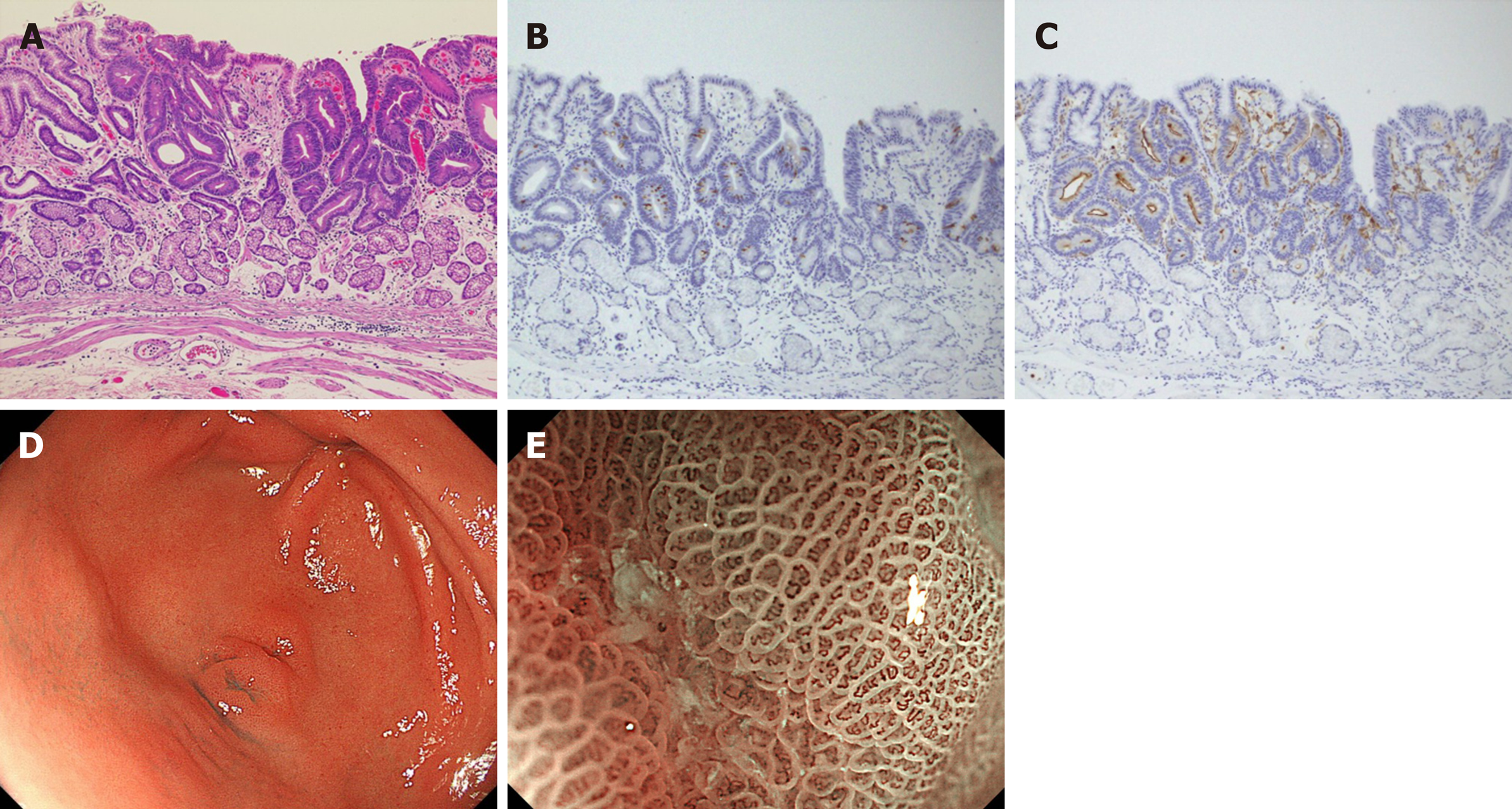
Figure 3 Intestinal-type adenocarcinoma.
A: The tubular structures lined by tall columnar cells with hyperchromatic, pencillate, and pseudostratified nuclei, hematoxylin and eosin, original magnification 4 ×; B: MUC2 was weakly positive for cancer cells; C: CD10 was positive for cancer cells; D: 0-IIa+IIc type tumor mimicking verrucosa on the gastric antrum, white light endoscopy; E: An irregular microvascular pattern and irregular microsurface pattern with a demarcation line were observed just in the recessed area, magnifying endoscopy with narrow-band imaging.
- Citation: Sato C, Hirasawa K, Tateishi Y, Ozeki Y, Sawada A, Ikeda R, Fukuchi T, Nishio M, Kobayashi R, Makazu M, Kaneko H, Inayama Y, Maeda S. Clinicopathological features of early gastric cancers arising in Helicobacter pylori uninfected patients. World J Gastroenterol 2020; 26(20): 2618-2631
- URL: https://www.wjgnet.com/1007-9327/full/v26/i20/2618.htm
- DOI: https://dx.doi.org/10.3748/wjg.v26.i20.2618


