Copyright
©The Author(s) 2020.
World J Gastroenterol. May 21, 2020; 26(19): 2403-2415
Published online May 21, 2020. doi: 10.3748/wjg.v26.i19.2403
Published online May 21, 2020. doi: 10.3748/wjg.v26.i19.2403
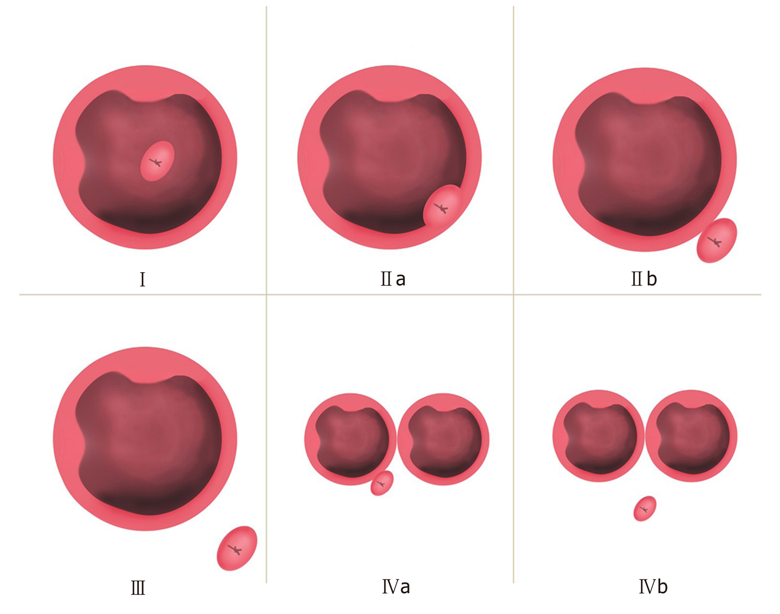
Figure 2 Li-Tanaka classification of periampullary diverticulum.
Type I: The papilla is located inside the diverticulum and not adjacent to the margin; type II: The papilla is located in the margin of the diverticulum (type IIa: Inside of the margin; type IIb: Outside of the margin, < 1 cm); type III: The papilla is located outside the margin, ≥ 1 cm; type IV: The papilla is located in the margin of the diverticulum, ≥ 2 diverticula (type IVa: The papilla is located outside the margins of at least one diverticulum, < 1 cm; type IVb: The papilla is located outside the margins of all the diverticula, ≥ 1 cm).
- Citation: Yue P, Zhu KX, Wang HP, Meng WB, Liu JK, Zhang L, Zhu XL, Zhang H, Miao L, Wang ZF, Zhou WC, Suzuki A, Tanaka K, Li X. Clinical significance of different periampullary diverticulum classifications for endoscopic retrograde cholangiopancreatography cannulation. World J Gastroenterol 2020; 26(19): 2403-2415
- URL: https://www.wjgnet.com/1007-9327/full/v26/i19/2403.htm
- DOI: https://dx.doi.org/10.3748/wjg.v26.i19.2403


