Copyright
©The Author(s) 2020.
World J Gastroenterol. May 21, 2020; 26(19): 2276-2285
Published online May 21, 2020. doi: 10.3748/wjg.v26.i19.2276
Published online May 21, 2020. doi: 10.3748/wjg.v26.i19.2276
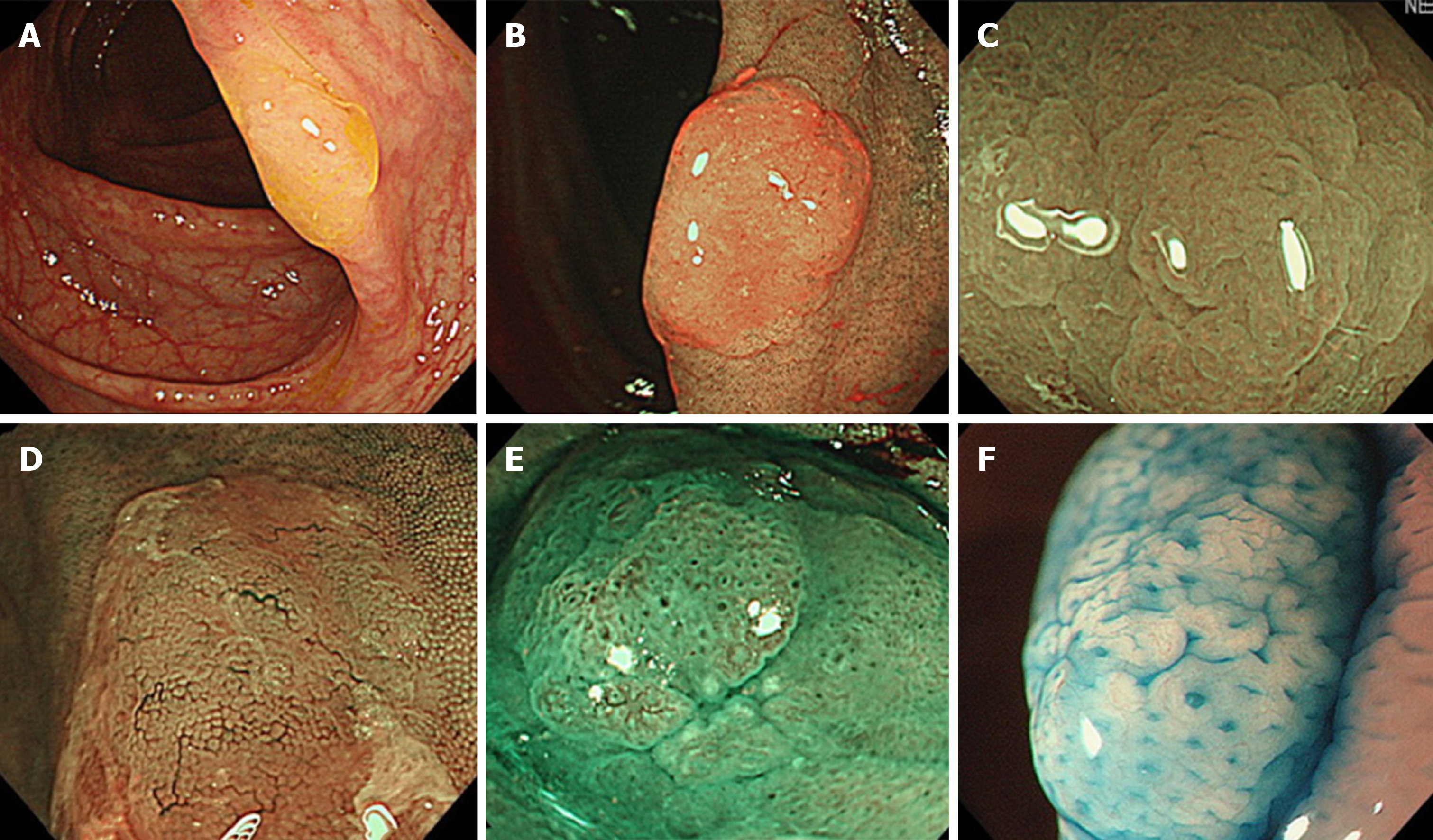
Figure 2 Endoscopic features of sessile serrated lesions.
A: Mucous cap (white light endoscopy); B: Red cap sign [narrow-band imaging (NBI) endoscopy]; C: Cloud-like surface (white light or NBI endoscopy); D: Dilated and branching vessels (NBI endoscopy); E: Expanded crypt openings (NBI endoscopy); F: Type II open-shape pits (chromoendoscopy).
- Citation: Sano W, Hirata D, Teramoto A, Iwatate M, Hattori S, Fujita M, Sano Y. Serrated polyps of the colon and rectum: Remove or not? World J Gastroenterol 2020; 26(19): 2276-2285
- URL: https://www.wjgnet.com/1007-9327/full/v26/i19/2276.htm
- DOI: https://dx.doi.org/10.3748/wjg.v26.i19.2276


