Copyright
©The Author(s) 2020.
World J Gastroenterol. May 14, 2020; 26(18): 2268-2275
Published online May 14, 2020. doi: 10.3748/wjg.v26.i18.2268
Published online May 14, 2020. doi: 10.3748/wjg.v26.i18.2268
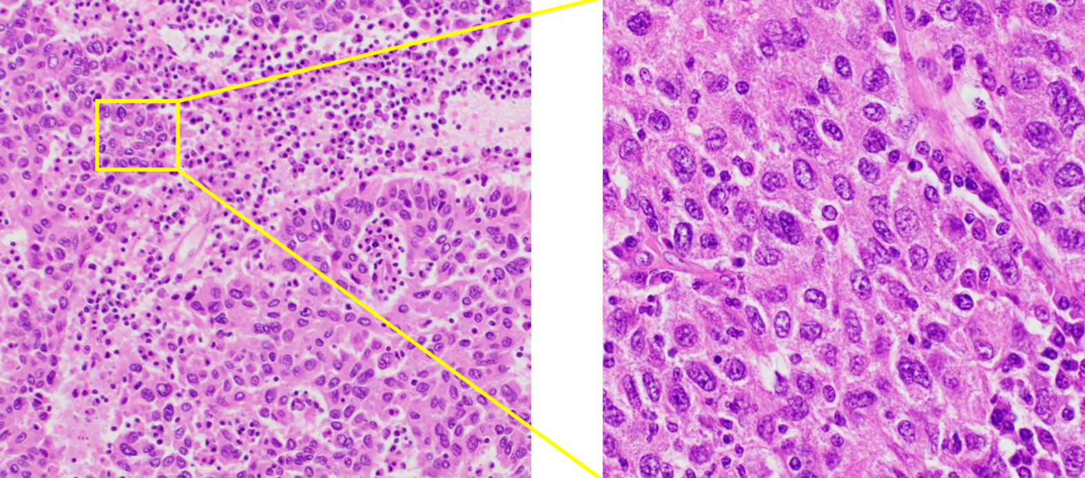
Figure 5 The microscopic examination of the tumor.
Microscopic examination of the tumor confirmed poorly differentiated carcinoma morphologically similar to the tumor biopsy by endoscopic ultrasound-guided fine needle aspiration Hematoxylin and eosin stain, magnification × 100 (left) and × 400 (right).
- Citation: Adachi Y, Hayashi H, Yusa T, Takematsu T, Matsumura K, Higashi T, Yamamura K, Yamao T, Imai K, Yamashita Y, Baba H. Ectopic hepatocellular carcinoma mimicking a retroperitoneal tumor: A case report. World J Gastroenterol 2020; 26(18): 2268-2275
- URL: https://www.wjgnet.com/1007-9327/full/v26/i18/2268.htm
- DOI: https://dx.doi.org/10.3748/wjg.v26.i18.2268


