Copyright
©The Author(s) 2020.
World J Gastroenterol. May 7, 2020; 26(17): 2111-2118
Published online May 7, 2020. doi: 10.3748/wjg.v26.i17.2111
Published online May 7, 2020. doi: 10.3748/wjg.v26.i17.2111
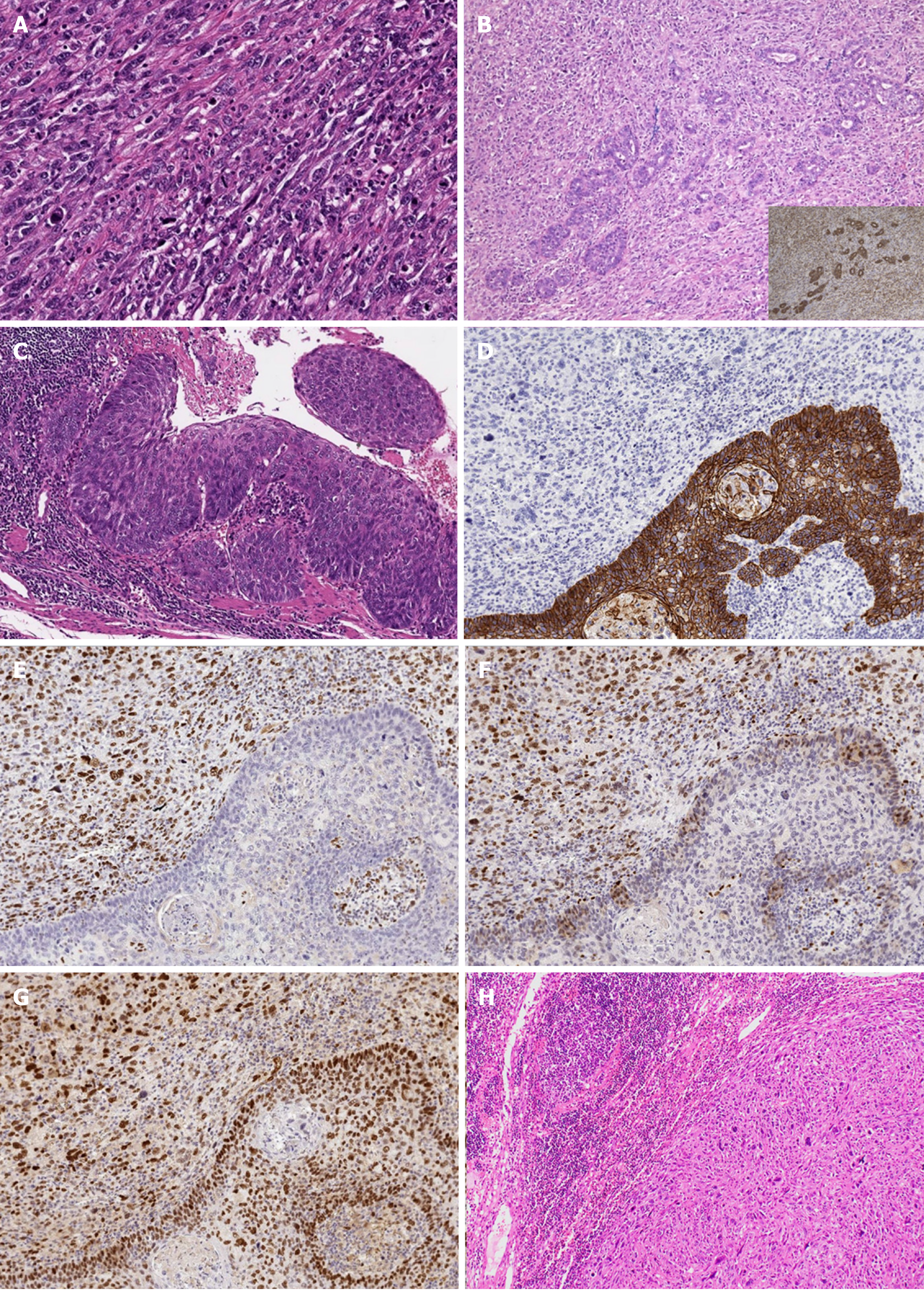
Figure 4 Histopathological and immunohistochemical findings.
A: Dense proliferation and infiltration of spindle cells mixed with giant cells with large atypical nuclei and polynuclear cells were observed (hematoxylin–eosin staining, × 200); B: Adeno-carcinomatous components were detected in a section near the root (hematoxylin–eosin staining, × 100; inserted figure: immunohistochemical expression of CAM 5.2, × 100); C: Squamous cell carcinoma in situ was found in the mucosa of the basal part of the tumor (hematoxylin–eosin staining, × 100); D: E-cadherin was not detected in spindle cells but was detected in the regions of squamous cell carcinoma (× 100); E-G: Epithelial-mesenchymal transition-related markers, zinc finger E-box-binding homeobox 1 (E, × 100), TWIST (F, × 100), and snail family transcriptional repressor 2 (G, × 100) were detected in spindle cells; H: Lymph node metastases with sarcomatous components (hematoxylin–eosin staining, × 40).
- Citation: Okamoto H, Kikuchi H, Naganuma H, Kamei T. Multiple carcinosarcomas of the esophagus with adeno-carcinomatous components: A case report. World J Gastroenterol 2020; 26(17): 2111-2118
- URL: https://www.wjgnet.com/1007-9327/full/v26/i17/2111.htm
- DOI: https://dx.doi.org/10.3748/wjg.v26.i17.2111


