Copyright
©The Author(s) 2020.
World J Gastroenterol. May 7, 2020; 26(17): 2111-2118
Published online May 7, 2020. doi: 10.3748/wjg.v26.i17.2111
Published online May 7, 2020. doi: 10.3748/wjg.v26.i17.2111
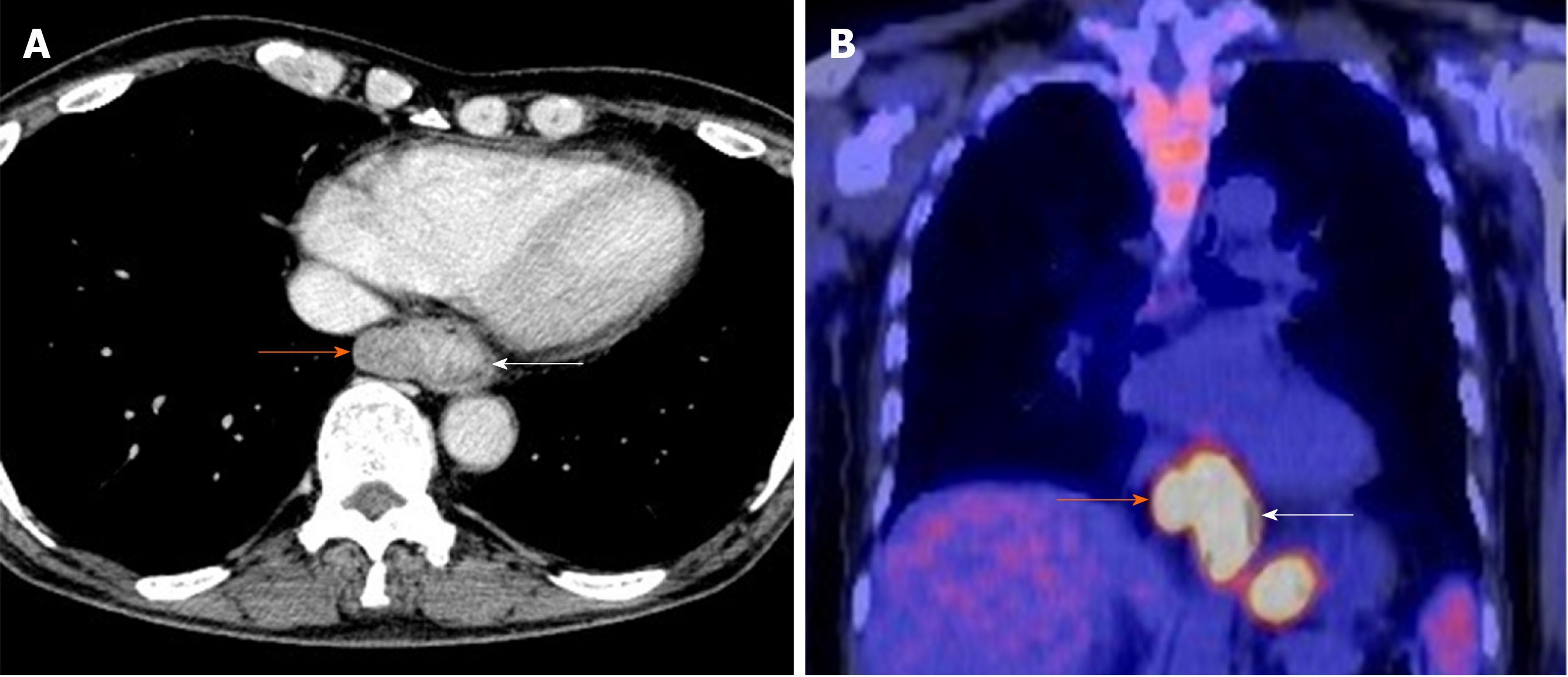
Figure 2 Enhanced computerized tomography and 18F-2-fluoro-2-deoxy-D-glucose positron-emission tomography.
A: Enhanced computerized tomography; B: 18F-2-fluoro-2-deoxy-D-glucose positron-emission tomography. The major axis of the main tumor was 42 mm and maximum standardized uptake value of the lesion was 13.9 at the white arrow. There suspected paraesophageal lymph node metastasis is indicated by the orange arrow.
- Citation: Okamoto H, Kikuchi H, Naganuma H, Kamei T. Multiple carcinosarcomas of the esophagus with adeno-carcinomatous components: A case report. World J Gastroenterol 2020; 26(17): 2111-2118
- URL: https://www.wjgnet.com/1007-9327/full/v26/i17/2111.htm
- DOI: https://dx.doi.org/10.3748/wjg.v26.i17.2111


