Copyright
©The Author(s) 2020.
World J Gastroenterol. May 7, 2020; 26(17): 2012-2029
Published online May 7, 2020. doi: 10.3748/wjg.v26.i17.2012
Published online May 7, 2020. doi: 10.3748/wjg.v26.i17.2012
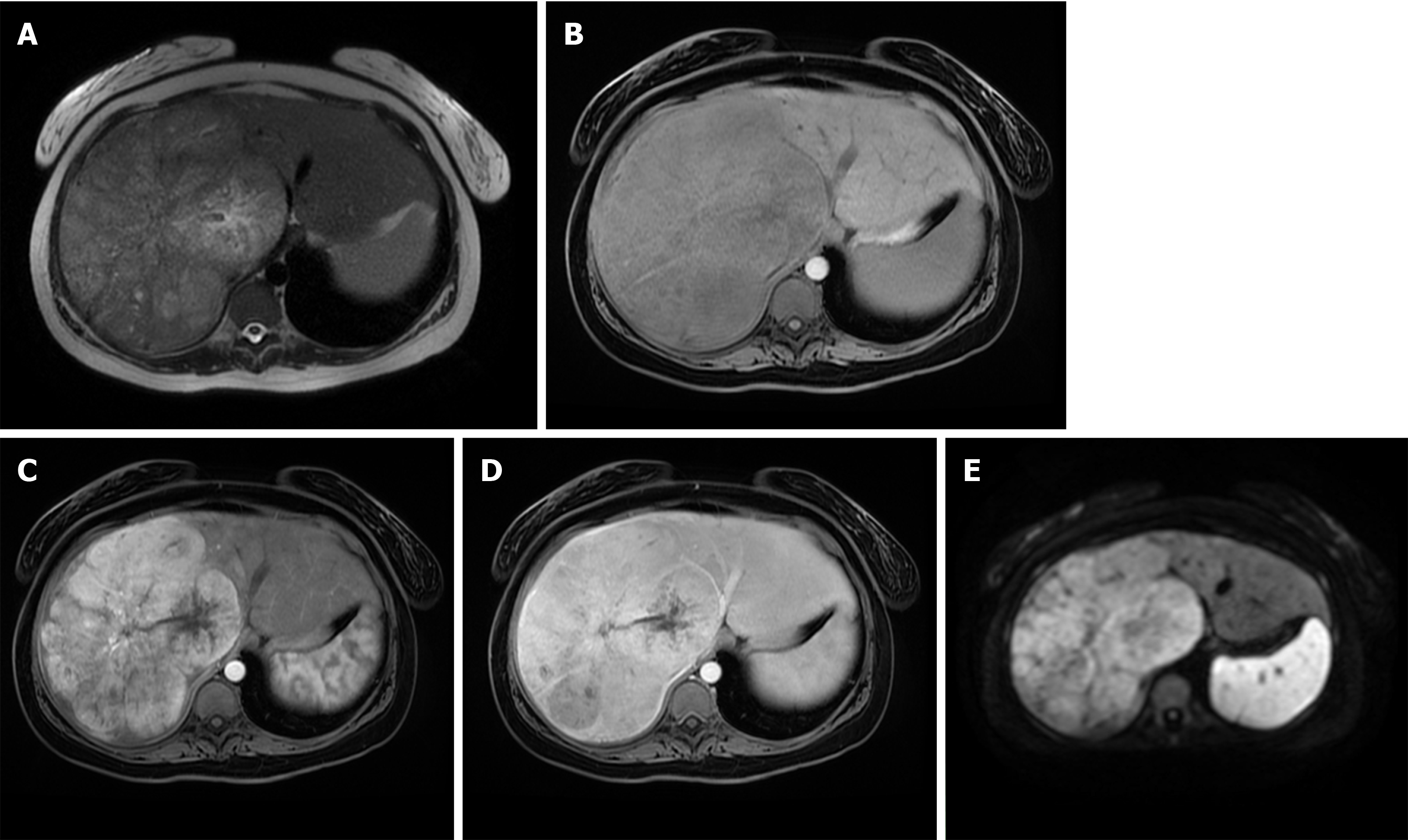
Figure 8 Fibrolamellar hepatocellular carcinoma in 23-year old woman without chronic liver disease.
A: Axial T2-weighted image shows large heterogeneous mass occupying almost whole right liver lobe; B-E: The tumor is hypointense on T1-weghted FS image (B), hypervascular on arterial phase (C) with washout in parts of the lesion on portal venous phase (D) and diffusion restriction (E).
- Citation: Kovac JD, Milovanovic T, Dugalic V, Dumic I. Pearls and pitfalls in magnetic resonance imaging of hepatocellular carcinoma. World J Gastroenterol 2020; 26(17): 2012-2029
- URL: https://www.wjgnet.com/1007-9327/full/v26/i17/2012.htm
- DOI: https://dx.doi.org/10.3748/wjg.v26.i17.2012


