Copyright
©The Author(s) 2020.
World J Gastroenterol. May 7, 2020; 26(17): 2012-2029
Published online May 7, 2020. doi: 10.3748/wjg.v26.i17.2012
Published online May 7, 2020. doi: 10.3748/wjg.v26.i17.2012
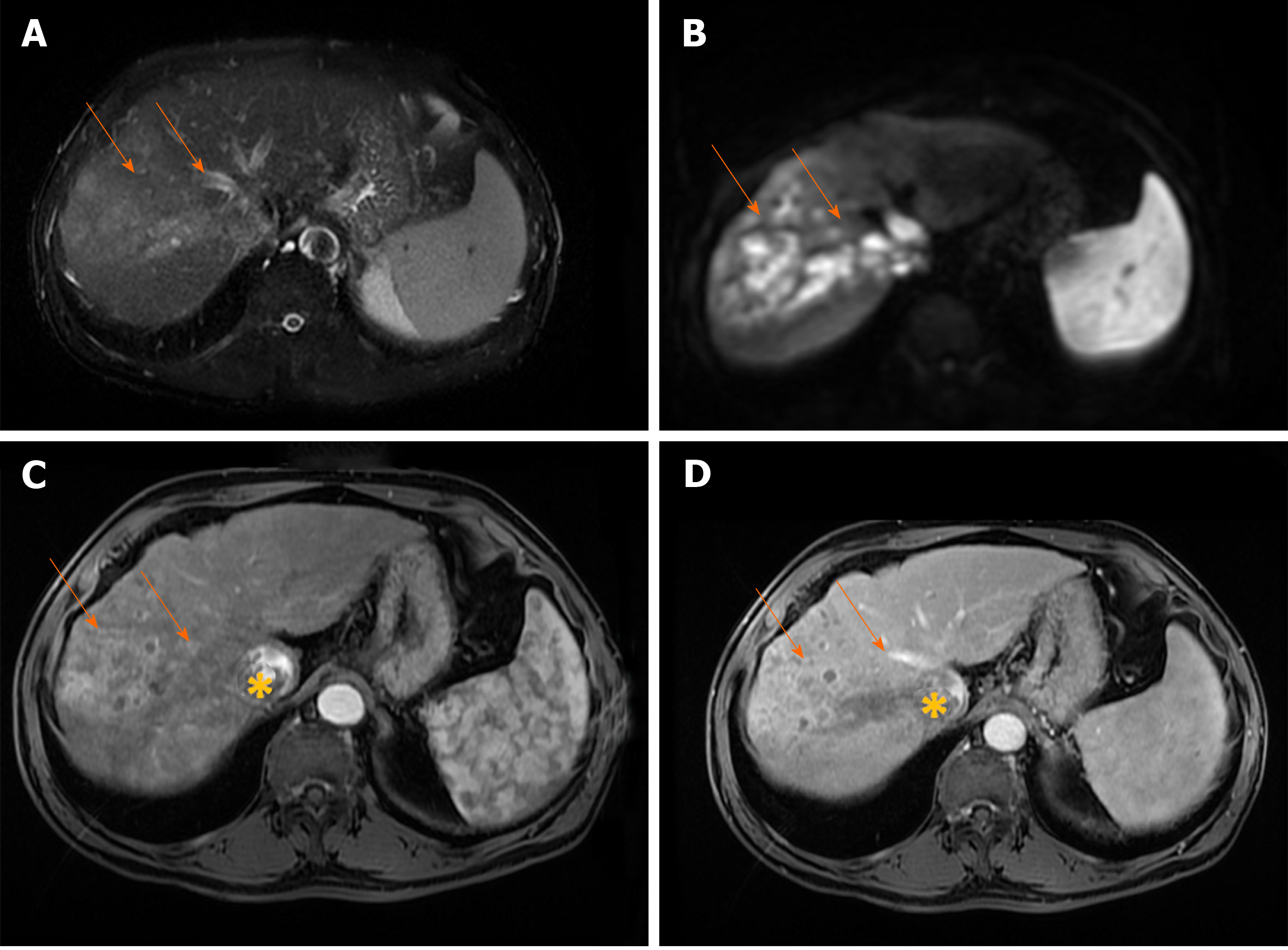
Figure 7 Infiltrative hepatocellular carcinoma in 71-year old man with cirrhosis.
A: Axial T2-weighted FS image shows ill-defined mass (arrows) in segments VII and VIII; B: On diffusion weighted image the lesion is hyperintense; C and D: The tumor is heterogeneously hyperintense on arterial phase (C), with washout in portal vein phase (D). Note also right hepatic vein thrombosis with propagation of the tumor thrombus in vena cava inferior (asterix).
- Citation: Kovac JD, Milovanovic T, Dugalic V, Dumic I. Pearls and pitfalls in magnetic resonance imaging of hepatocellular carcinoma. World J Gastroenterol 2020; 26(17): 2012-2029
- URL: https://www.wjgnet.com/1007-9327/full/v26/i17/2012.htm
- DOI: https://dx.doi.org/10.3748/wjg.v26.i17.2012


