Copyright
©The Author(s) 2020.
World J Gastroenterol. May 7, 2020; 26(17): 2012-2029
Published online May 7, 2020. doi: 10.3748/wjg.v26.i17.2012
Published online May 7, 2020. doi: 10.3748/wjg.v26.i17.2012
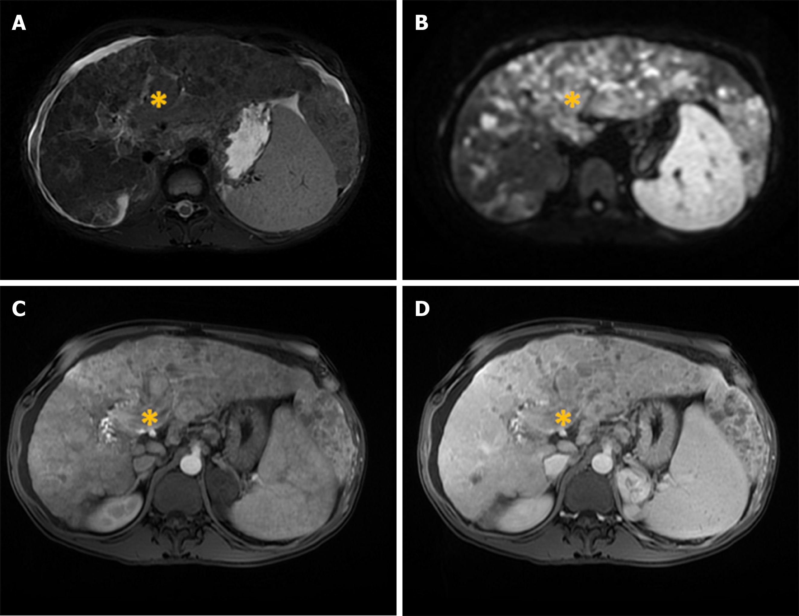
Figure 6 Diffuse hepatocellular carcinoma in 38-year-old man with long-standing Wilson disease.
Axial T2-weighted FS image shows multiple moderately hyperintense nodules scattered throughout liver parenchyma. A: Note also portal vein thrombosis with signal intensity of the thrombus similar to tumor nodules in the liver; B: On diffusion weighted image diffuse hyperintensity is seen corresponding to multiple tumor nodules; C and D: On arterial phase patchy enhancement can be seen including portal vein thrombus (C), with heterogeneous washout in portal vein phase (D).
- Citation: Kovac JD, Milovanovic T, Dugalic V, Dumic I. Pearls and pitfalls in magnetic resonance imaging of hepatocellular carcinoma. World J Gastroenterol 2020; 26(17): 2012-2029
- URL: https://www.wjgnet.com/1007-9327/full/v26/i17/2012.htm
- DOI: https://dx.doi.org/10.3748/wjg.v26.i17.2012


