Copyright
©The Author(s) 2020.
World J Gastroenterol. Apr 14, 2020; 26(14): 1601-1612
Published online Apr 14, 2020. doi: 10.3748/wjg.v26.i14.1601
Published online Apr 14, 2020. doi: 10.3748/wjg.v26.i14.1601
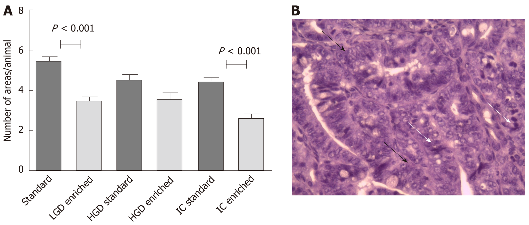
Figure 2 Results summarize and an explanatory example of intestinal carcinoma histological picture.
A: Mean number ± SD/mouse of microscopic areas of low-grade dysplasia, high grade dysplasia and intestinal carcinoma. B: A histological picture of intestinal carcinoma (hematoxylin-eosin stain) showing cell crowding and pleomorphism (white arrow), architectural loss and nuclear hyperchromatism (black arrow). LGD: Low-grade dysplasia; HGD: High grade dysplasia; IC: Intestinal carcinoma.
- Citation: Girardi B, Pricci M, Giorgio F, Piazzolla M, Iannone A, Losurdo G, Principi M, Barone M, Ierardi E, Di Leo A. Silymarin, boswellic acid and curcumin enriched dietetic formulation reduces the growth of inherited intestinal polyps in an animal model. World J Gastroenterol 2020; 26(14): 1601-1612
- URL: https://www.wjgnet.com/1007-9327/full/v26/i14/1601.htm
- DOI: https://dx.doi.org/10.3748/wjg.v26.i14.1601


