Copyright
©The Author(s) 2020.
World J Gastroenterol. Apr 7, 2020; 26(13): 1450-1462
Published online Apr 7, 2020. doi: 10.3748/wjg.v26.i13.1450
Published online Apr 7, 2020. doi: 10.3748/wjg.v26.i13.1450
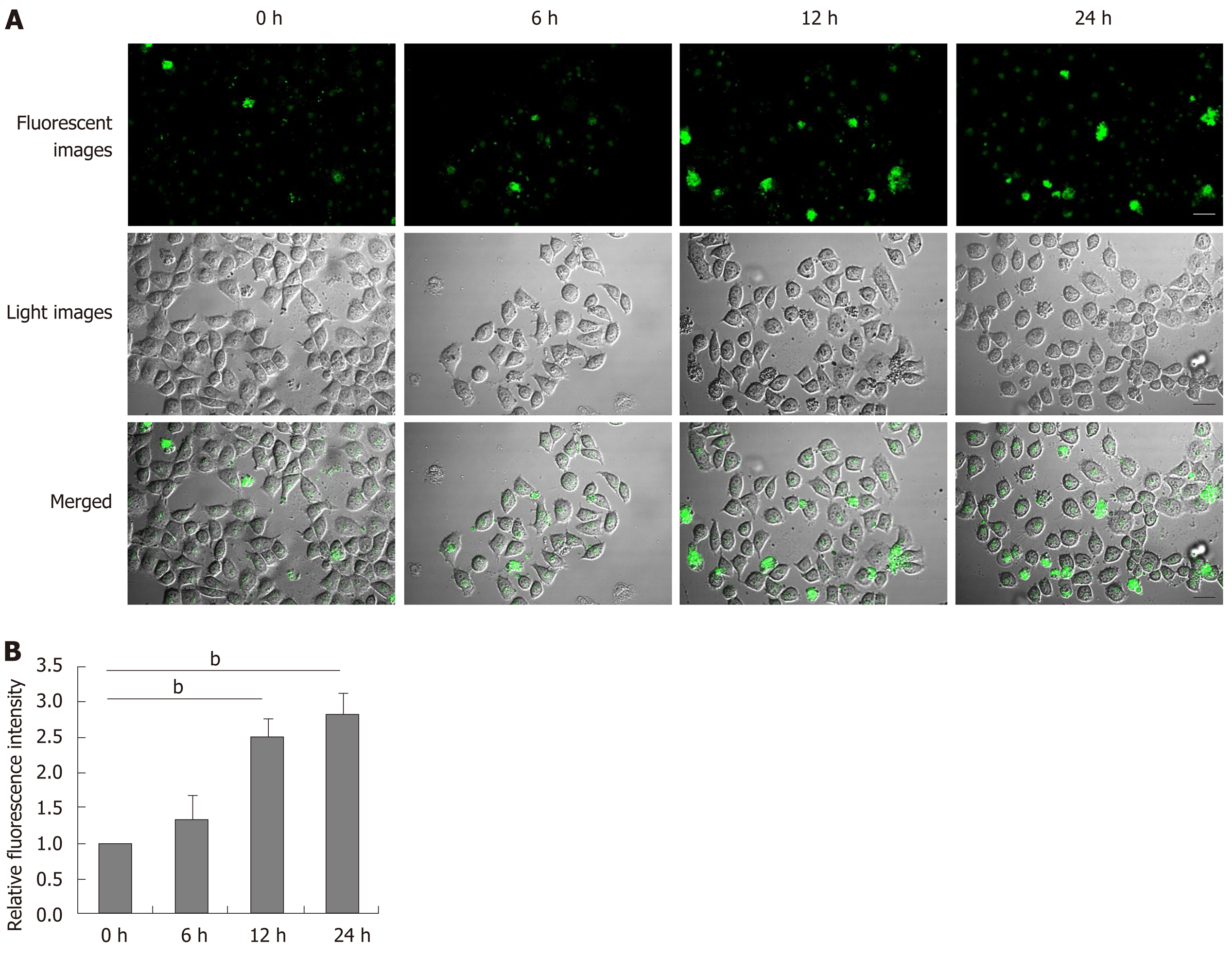
Figure 4 Dithiothreitol increases the levels of intracellular Ca2+ in BRL-3A cells.
After treatment with 2.0 mmol/L dithiothreitol for varying time periods, the cells were labeled by Fluo-3 AM. The cell morphology and fluorescent signals in cells were observed by microscopy. A: Representative images of Fluo-3 fluorescence and morphology in BRL-3A cells following dithiothreitol treatment (Scale bars: 25 μm); B: Quantification of fluorescence intensity in BRL-3A cells. Data are representative images (magnification 200 ×) or expressed as the mean ± SD of each group of cells from three separate experiments. bP < 0.01.
- Citation: Xie RJ, Hu XX, Zheng L, Cai S, Chen YS, Yang Y, Yang T, Han B, Yang Q. Calpain-2 activity promotes aberrant endoplasmic reticulum stress-related apoptosis in hepatocytes. World J Gastroenterol 2020; 26(13): 1450-1462
- URL: https://www.wjgnet.com/1007-9327/full/v26/i13/1450.htm
- DOI: https://dx.doi.org/10.3748/wjg.v26.i13.1450


