Copyright
©The Author(s) 2020.
World J Gastroenterol. Apr 7, 2020; 26(13): 1439-1449
Published online Apr 7, 2020. doi: 10.3748/wjg.v26.i13.1439
Published online Apr 7, 2020. doi: 10.3748/wjg.v26.i13.1439
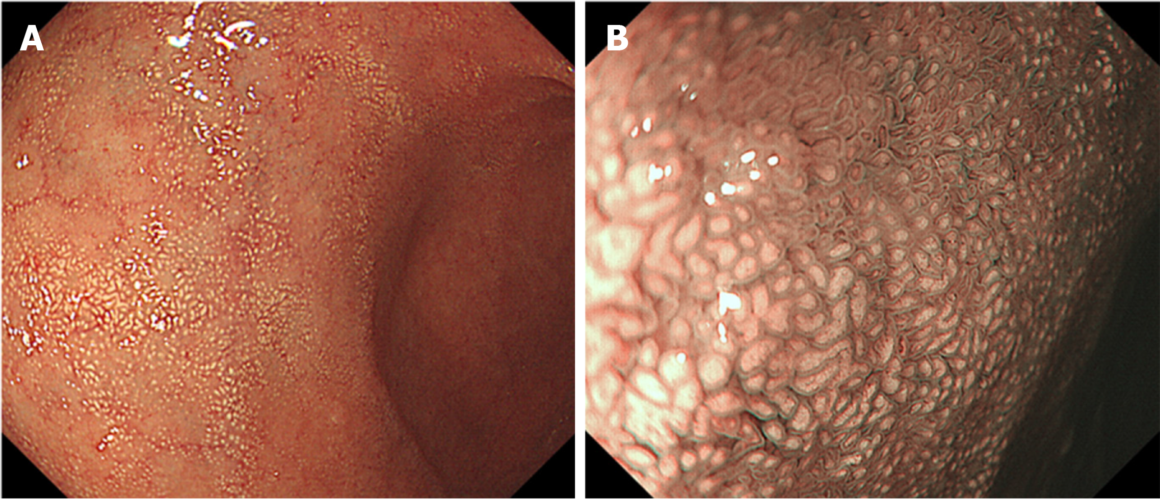
Figure 7 Endoscopic images of the duodenal lanthanum deposition.
A: A lanthanum-related lesion in the duodenum presents with white mucosa; B: Magnified image with narrow-band imaging shows white depositions within the duodenal villi.
- Citation: Iwamuro M, Urata H, Tanaka T, Okada H. Review of the diagnosis of gastrointestinal lanthanum deposition. World J Gastroenterol 2020; 26(13): 1439-1449
- URL: https://www.wjgnet.com/1007-9327/full/v26/i13/1439.htm
- DOI: https://dx.doi.org/10.3748/wjg.v26.i13.1439


