Copyright
©The Author(s) 2020.
World J Gastroenterol. Apr 7, 2020; 26(13): 1439-1449
Published online Apr 7, 2020. doi: 10.3748/wjg.v26.i13.1439
Published online Apr 7, 2020. doi: 10.3748/wjg.v26.i13.1439
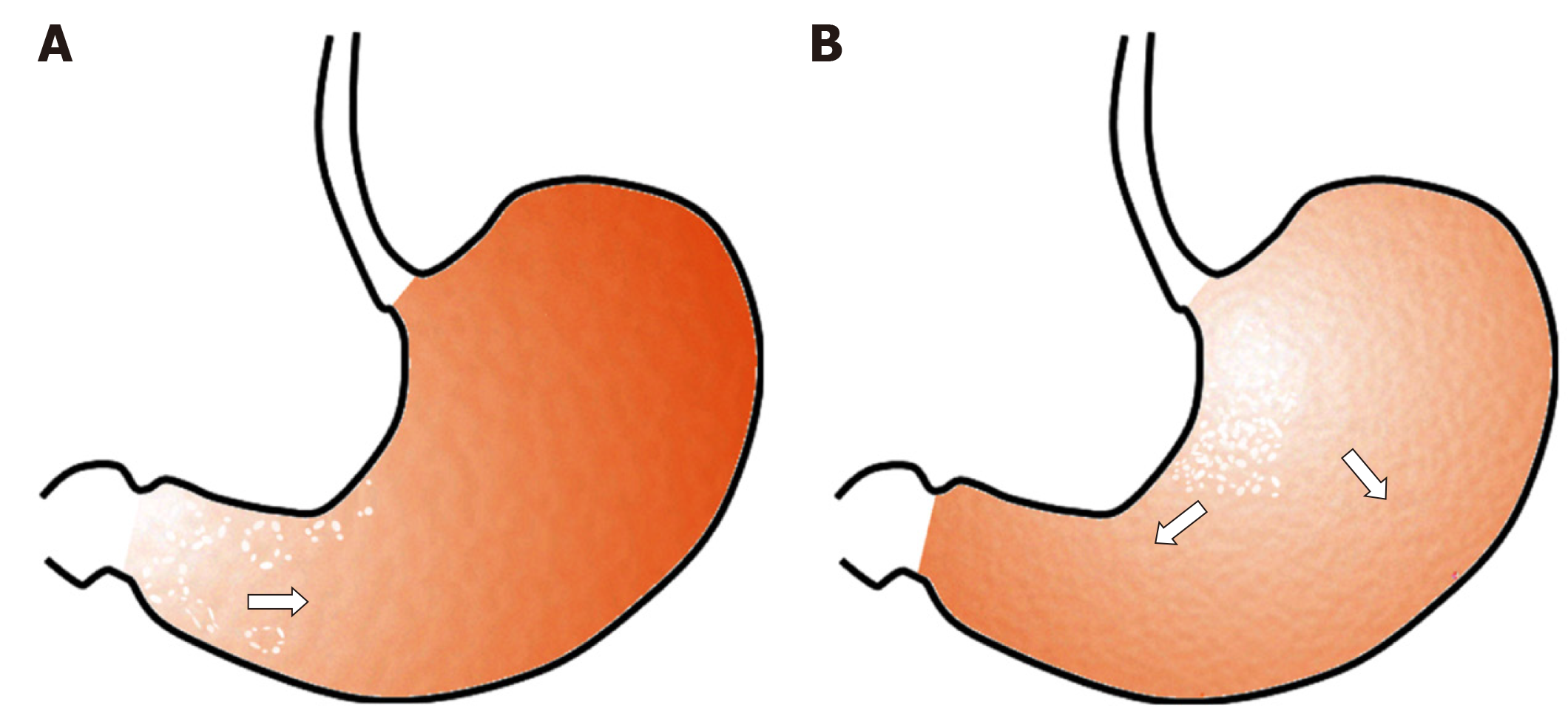
Figure 6 Hypothesis regarding the pattern of lanthanum deposition in the gastric mucosa with or without atrophy.
A: In the atrophic mucosa, particularly in areas with intestinal metaplasia, lanthanum deposition probably presents with annular and/or granular white lesions, predominantly in the gastric antrum and angle. As intestinal metaplasia expands, the size of the areas with lanthanum deposition may increase; B: In the gastric mucosa without atrophy, lanthanum primarily deposits in the lesser curvature and posterior wall of the gastric body and presents diffuse whitish lesions. The area of lanthanum deposition probably expands as time elapses unless the patient stops lanthanum carbonate.
- Citation: Iwamuro M, Urata H, Tanaka T, Okada H. Review of the diagnosis of gastrointestinal lanthanum deposition. World J Gastroenterol 2020; 26(13): 1439-1449
- URL: https://www.wjgnet.com/1007-9327/full/v26/i13/1439.htm
- DOI: https://dx.doi.org/10.3748/wjg.v26.i13.1439


