Copyright
©The Author(s) 2020.
World J Gastroenterol. Mar 21, 2020; 26(11): 1128-1141
Published online Mar 21, 2020. doi: 10.3748/wjg.v26.i11.1128
Published online Mar 21, 2020. doi: 10.3748/wjg.v26.i11.1128
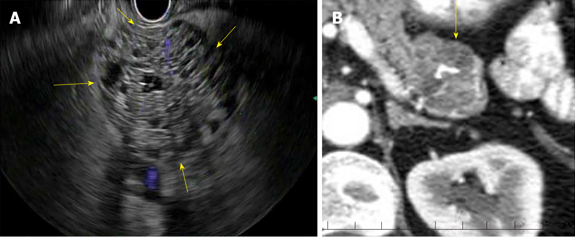
Figure 1 Endoscopic ultrasound (Honeycomb appearance).
A: Endoscopic ultrasound: Serous cystadenoma; B: Computed tomography: Serous Cystadenoma. Computed tomography: Portal venous phase showing calcification.
- Citation: Lanke G, Lee JH. Similarities and differences in guidelines for the management of pancreatic cysts. World J Gastroenterol 2020; 26(11): 1128-1141
- URL: https://www.wjgnet.com/1007-9327/full/v26/i11/1128.htm
- DOI: https://dx.doi.org/10.3748/wjg.v26.i11.1128


