Copyright
©The Author(s) 2019.
World J Gastroenterol. Mar 7, 2019; 25(9): 1100-1115
Published online Mar 7, 2019. doi: 10.3748/wjg.v25.i9.1100
Published online Mar 7, 2019. doi: 10.3748/wjg.v25.i9.1100
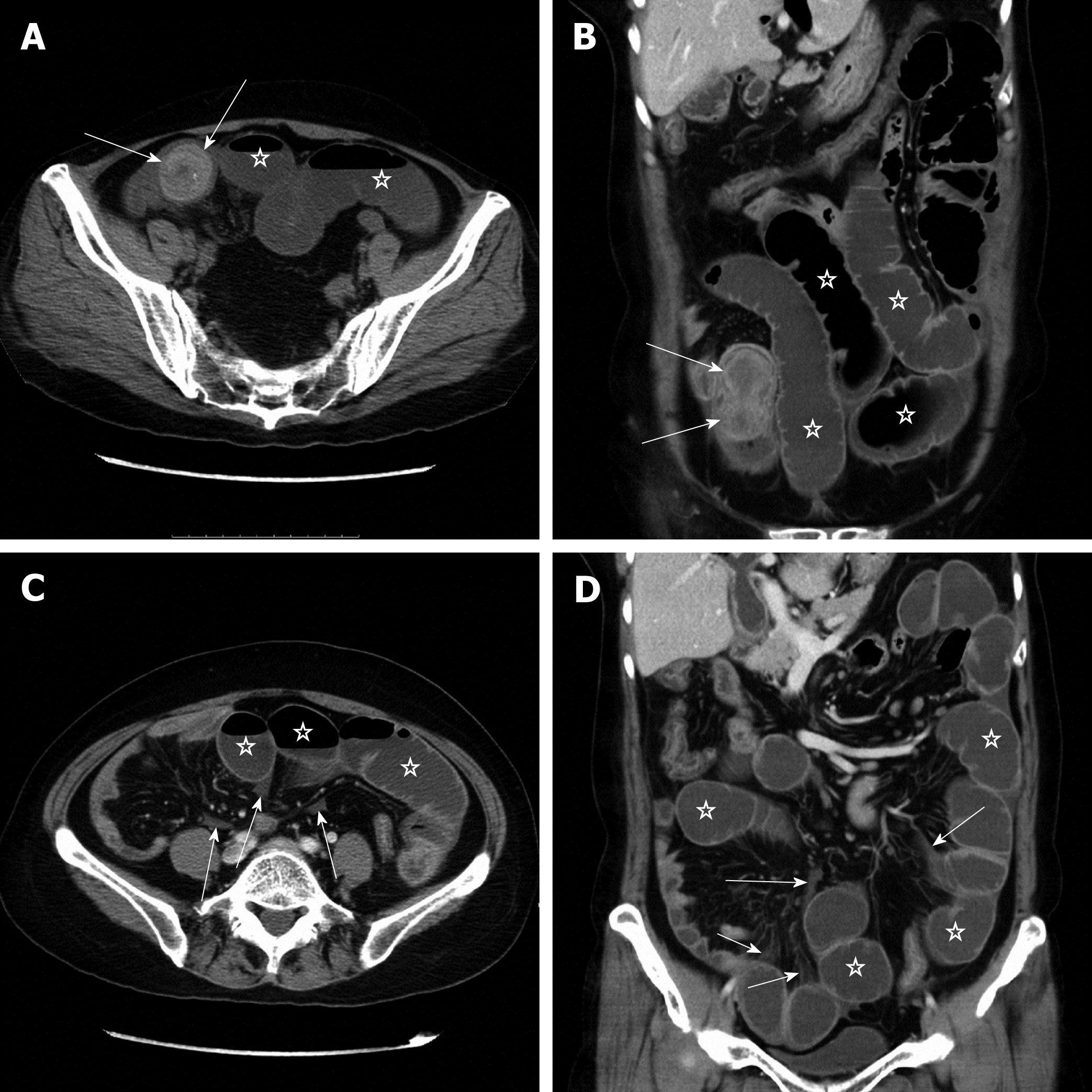
Figure 3 Protocol 3 applied in a 57-year-old woman with a bezoar causing small bowel obstruction.
Because all assessment parameters could be evaluated with high self-confidence and satisfaction using the conventional axial and coronal reformations, the reader did not perform the multiple post-processing techniques. A and B: Multidetector computed tomography (MDCT) axial and coronal images of the abdomen reveal a well-defined, dumbbell-shaped bezoar (arrows) with a size of 5.8 cm × 3.5 cm × 3.5 cm in the distal segment of the small bowel. The bowel loops (pentagram) proximal to the bezoar are severely dilated with a maximum diameter of 4.5 cm. C and D: Additional MDCT axial and coronal images indicate small patchy mesenteric haziness and fluid (arrows), as well as the dilated bowel loops (pentagram), but no more signs of secondary bowel ischemia were found in the patient.
- Citation: Kuang LQ, Tang W, Li R, Cheng C, Tang SY, Wang Y. Optimized protocol of multiple post-processing techniques improves diagnostic accuracy of multidetector computed tomography in assessment of small bowel obstruction compared with conventional axial and coronal reformations. World J Gastroenterol 2019; 25(9): 1100-1115
- URL: https://www.wjgnet.com/1007-9327/full/v25/i9/1100.htm
- DOI: https://dx.doi.org/10.3748/wjg.v25.i9.1100


