Copyright
©The Author(s) 2019.
World J Gastroenterol. Feb 21, 2019; 25(7): 870-879
Published online Feb 21, 2019. doi: 10.3748/wjg.v25.i7.870
Published online Feb 21, 2019. doi: 10.3748/wjg.v25.i7.870
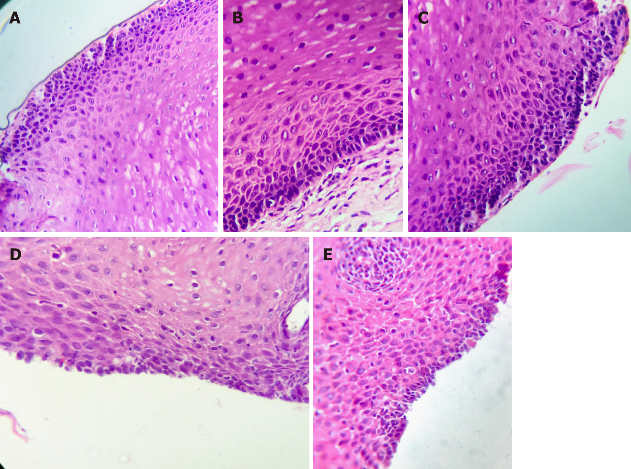
Figure 3 Representative photomicrograph showing low grade squamous dysplasia: The basal one third of the epithelium shows epithelial cell disorganization, nuclear pleomorphism, hyperchromasia, and cellular crowding (× 400), patient 1 (A-C), patient 2 (D and E).
- Citation: Eskander A, Ghobrial C, Mohsen NA, Mounir B, Abd EL-Kareem D, Tarek S, El-Shabrawi MH. Histopathological changes in the oesophageal mucosa in Egyptian children with corrosive strictures: A single-centre vast experience. World J Gastroenterol 2019; 25(7): 870-879
- URL: https://www.wjgnet.com/1007-9327/full/v25/i7/870.htm
- DOI: https://dx.doi.org/10.3748/wjg.v25.i7.870


