Copyright
©The Author(s) 2019.
World J Gastroenterol. Feb 21, 2019; 25(7): 744-776
Published online Feb 21, 2019. doi: 10.3748/wjg.v25.i7.744
Published online Feb 21, 2019. doi: 10.3748/wjg.v25.i7.744
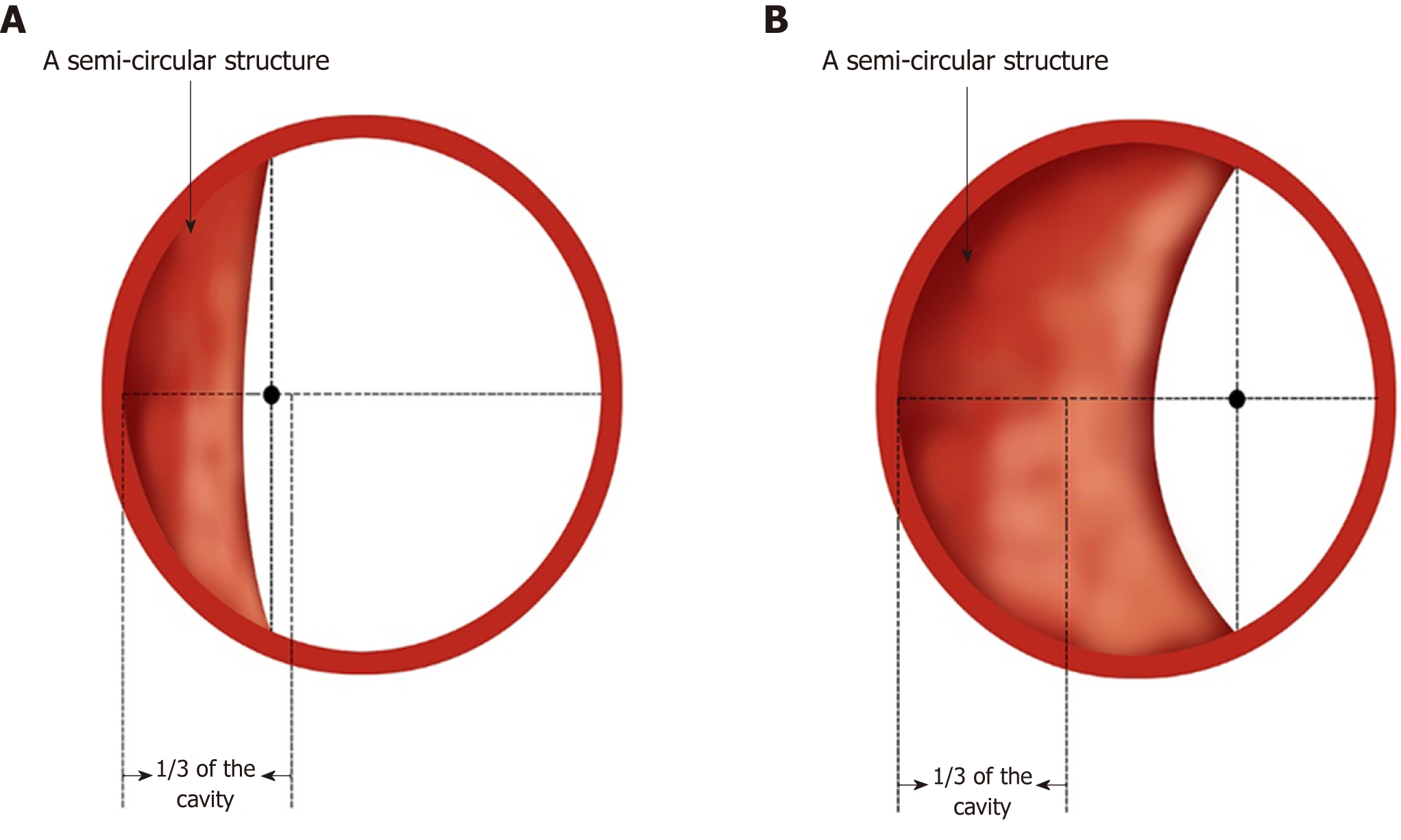
Figure 4 Simulated diagram of endoscopic observations in Ling IIb and Ling IIc.
A: Ling IIb. The arrows indicate 1/3 of the oesophageal cavity, and the semi-annular structure’s midpoint remains within this range; B: Ling IIc. The arrows indicate 1/3 of the oesophageal cavity, and the crescent-like structure's midpoint goes beyond this range.
- Citation: Chai NL, Li HK, Linghu EQ, Li ZS, Zhang ST, Bao Y, Chen WG, Chiu PW, Dang T, Gong W, Han ST, Hao JY, He SX, Hu B, Hu B, Huang XJ, Huang YH, Jin ZD, Khashab MA, Lau J, Li P, Li R, Liu DL, Liu HF, Liu J, Liu XG, Liu ZG, Ma YC, Peng GY, Rong L, Sha WH, Sharma P, Sheng JQ, Shi SS, Seo DW, Sun SY, Wang GQ, Wang W, Wu Q, Xu H, Xu MD, Yang AM, Yao F, Yu HG, Zhou PH, Zhang B, Zhang XF, Zhai YQ. Consensus on the digestive endoscopic tunnel technique. World J Gastroenterol 2019; 25(7): 744-776
- URL: https://www.wjgnet.com/1007-9327/full/v25/i7/744.htm
- DOI: https://dx.doi.org/10.3748/wjg.v25.i7.744


