Copyright
©The Author(s) 2019.
World J Gastroenterol. Feb 14, 2019; 25(6): 707-718
Published online Feb 14, 2019. doi: 10.3748/wjg.v25.i6.707
Published online Feb 14, 2019. doi: 10.3748/wjg.v25.i6.707
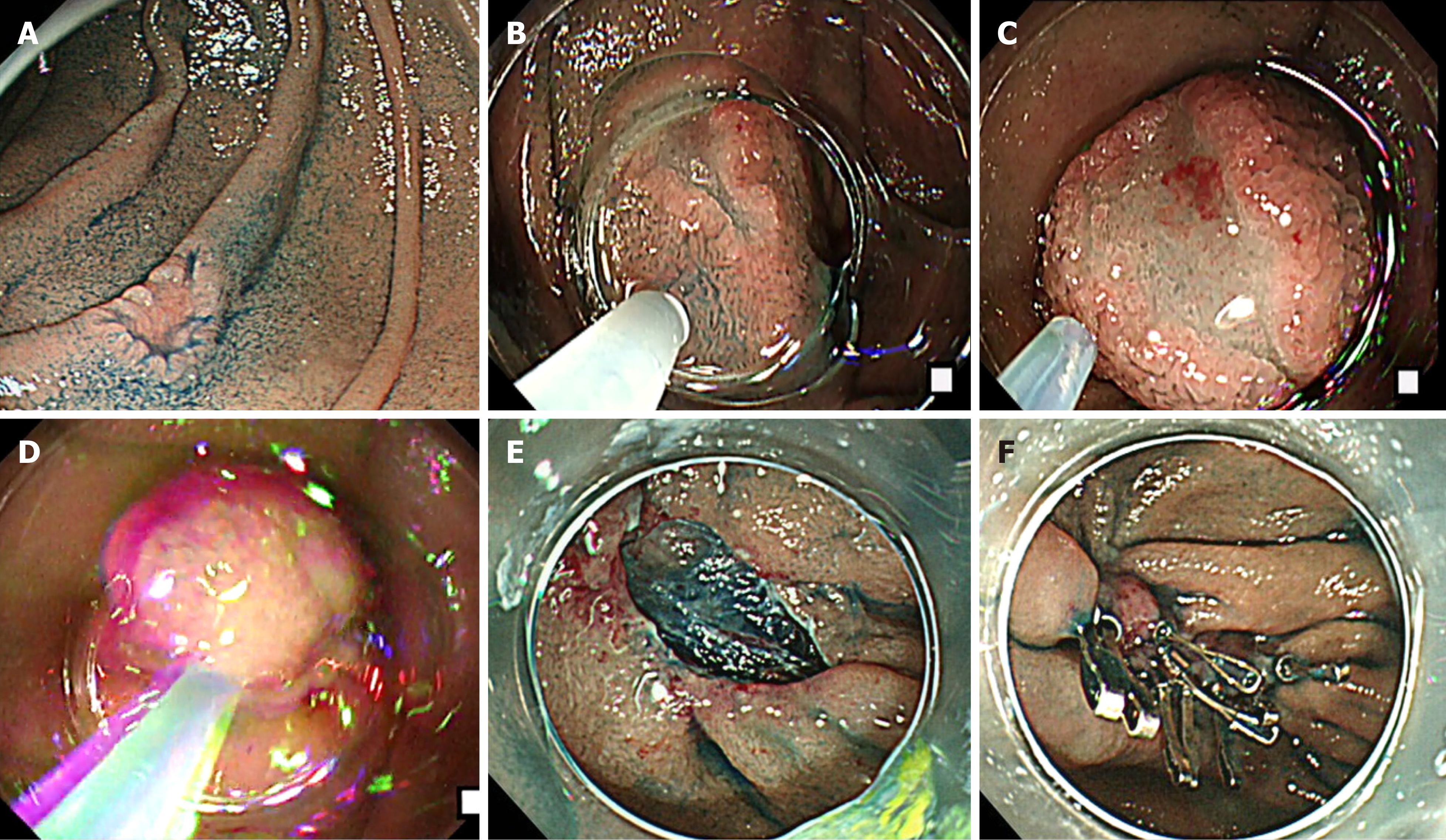
Figure 1 The cap-assisted endoscopic mucosal resection method of superficial non-ampullary duodenal epithelial tumor.
This is the “suck and shake” technique. Type 0-IIc lesion, 10 mm × 5 mm, mucosal carcinoma, cut-end negative. A: Indigo carmine spraying view. Depressed-type lesion was located in the second portion of the duodenum; B: Injecting the glycerol into submucosal layer; C: Sucking the lesion; D: Shaking the lesion to prevent muscle layer involvement; E: Ulcer findings just after lesion removal. The lesion was resected en bloc, and there was no bleeding or perforation in the ulcer just after the procedure; F: Closing the ulcer floor completely by clip to prevent delayed bleeding and perforation.
- Citation: Hara Y, Goda K, Dobashi A, Ohya TR, Kato M, Sumiyama K, Mitsuishi T, Hirooka S, Ikegami M, Tajiri H. Short- and long-term outcomes of endoscopically treated superficial non-ampullary duodenal epithelial tumors. World J Gastroenterol 2019; 25(6): 707-718
- URL: https://www.wjgnet.com/1007-9327/full/v25/i6/707.htm
- DOI: https://dx.doi.org/10.3748/wjg.v25.i6.707


