Copyright
©The Author(s) 2019.
World J Gastroenterol. Feb 14, 2019; 25(6): 644-658
Published online Feb 14, 2019. doi: 10.3748/wjg.v25.i6.644
Published online Feb 14, 2019. doi: 10.3748/wjg.v25.i6.644
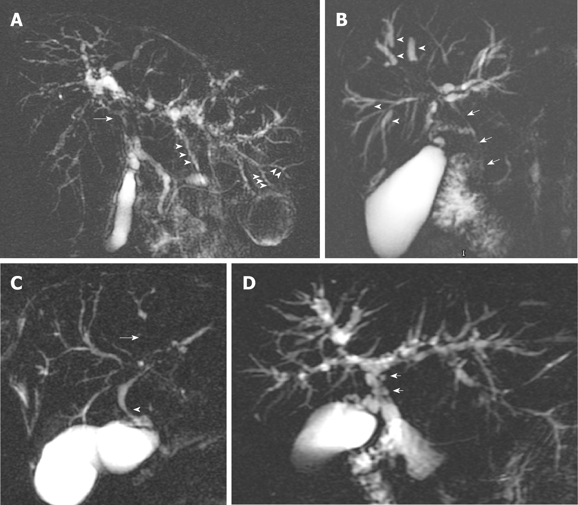
Figure 1 Magnetic resonance cholangiopancreatography annotating typical features of large-duct-primary sclerosing cholangitis.
A: Common hepatic duct stricture (arrow) and intrahepatic duct beading (arrowheads); B: Dominant stricture (arrow) and dilated proximal intrahepatic ducts secondary to distal strictures (arrowheads); C: Area of non-filling of intrahepatic ducts indicating tight stricture (arrow) and common hepatic duct stricture (arrowhead); D: Pseudodiverticula.
- Citation: Selvaraj EA, Culver EL, Bungay H, Bailey A, Chapman RW, Pavlides M. Evolving role of magnetic resonance techniques in primary sclerosing cholangitis. World J Gastroenterol 2019; 25(6): 644-658
- URL: https://www.wjgnet.com/1007-9327/full/v25/i6/644.htm
- DOI: https://dx.doi.org/10.3748/wjg.v25.i6.644


