Copyright
©The Author(s) 2019.
World J Gastroenterol. Feb 7, 2019; 25(5): 567-583
Published online Feb 7, 2019. doi: 10.3748/wjg.v25.i5.567
Published online Feb 7, 2019. doi: 10.3748/wjg.v25.i5.567
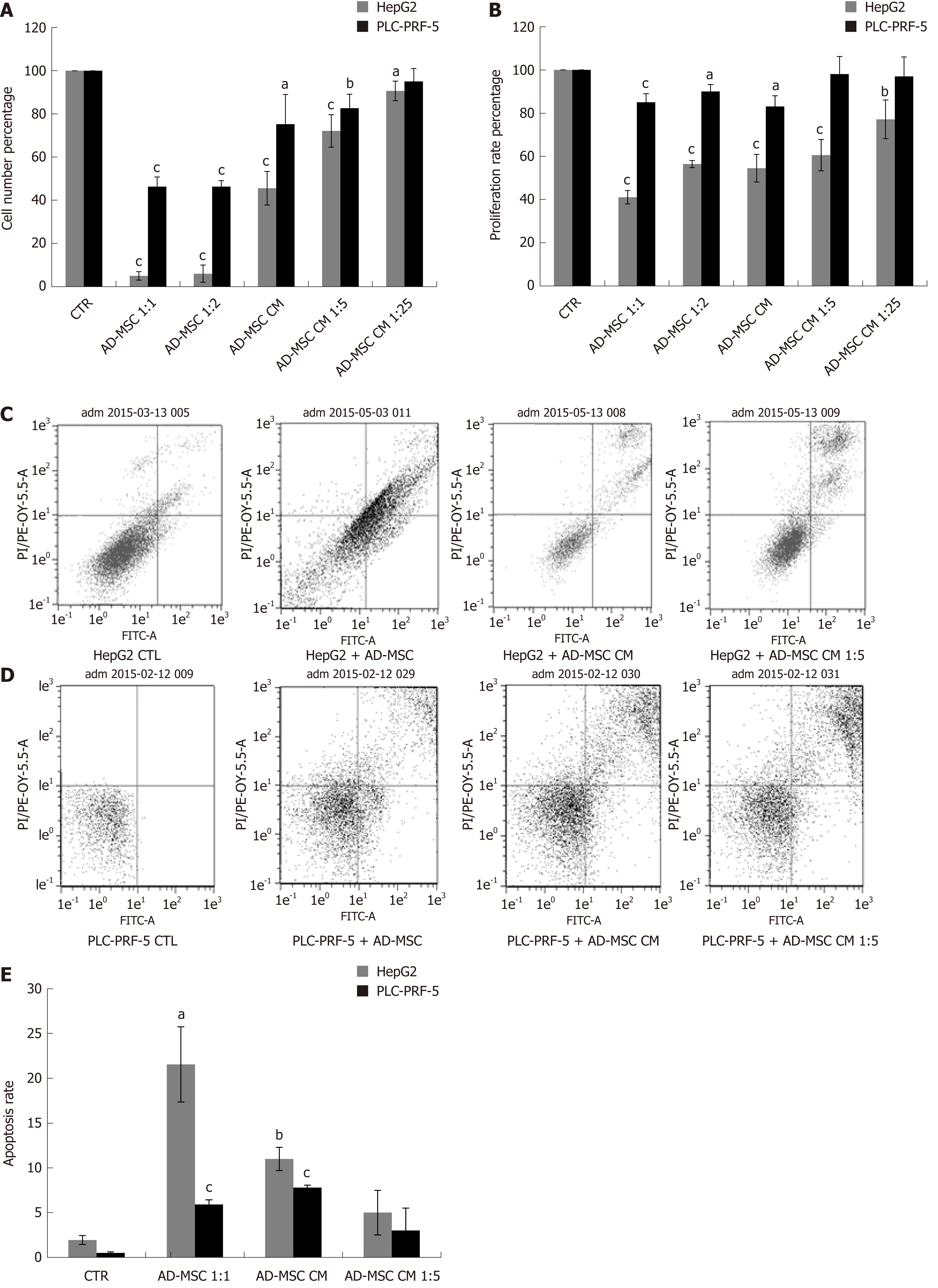
Figure 2 Effect of adipose-derived mesenchymal stem cells and their conditioned media on hepatocellular carcinoma cell proliferation and apoptosis.
HCC cells (2 × 10⁵) were seeded in six-well coculture plates in the presence or absence of ADMSCs and ADMSC CM, undiluted or diluted 1:5 or 1:25 for 48 h. The proliferation of HepG2 and PLC-PRF-5 HCC cell lines were measured by (A) cell count assay and (B) WST-8 proliferation tests; The apoptosis of HepG2 (C) and PLC-PRF (D) cells co-cultured as above was measured by flow cytometry using Annexin V/PI test kit; C and D: Two representative experiments of apoptosis in HepG2 and PLC-PRF cells, respectively; E: The average rate of apoptosis in HepG2 and PLC-PRF-5 cells induced by ADMSCs, ADMSC CM. Data are representative of three independent experiments, each repeated in triplicate. All data are represented as mean ± SD (aP < 0.05, bP < 0.01, cP < 0.001). CTR: Control; ADMSC: Adipose-derived mesenchymal stem cell; CM: Conditioned media; HCC: Hepatocellular carcinoma.
- Citation: Serhal R, Saliba N, Hilal G, Moussa M, Hassan GS, El Atat O, Alaaeddine N. Effect of adipose-derived mesenchymal stem cells on hepatocellular carcinoma: In vitro inhibition of carcinogenesis. World J Gastroenterol 2019; 25(5): 567-583
- URL: https://www.wjgnet.com/1007-9327/full/v25/i5/567.htm
- DOI: https://dx.doi.org/10.3748/wjg.v25.i5.567


