Copyright
©The Author(s) 2019.
World J Gastroenterol. Dec 7, 2019; 25(45): 6693-6703
Published online Dec 7, 2019. doi: 10.3748/wjg.v25.i45.6693
Published online Dec 7, 2019. doi: 10.3748/wjg.v25.i45.6693
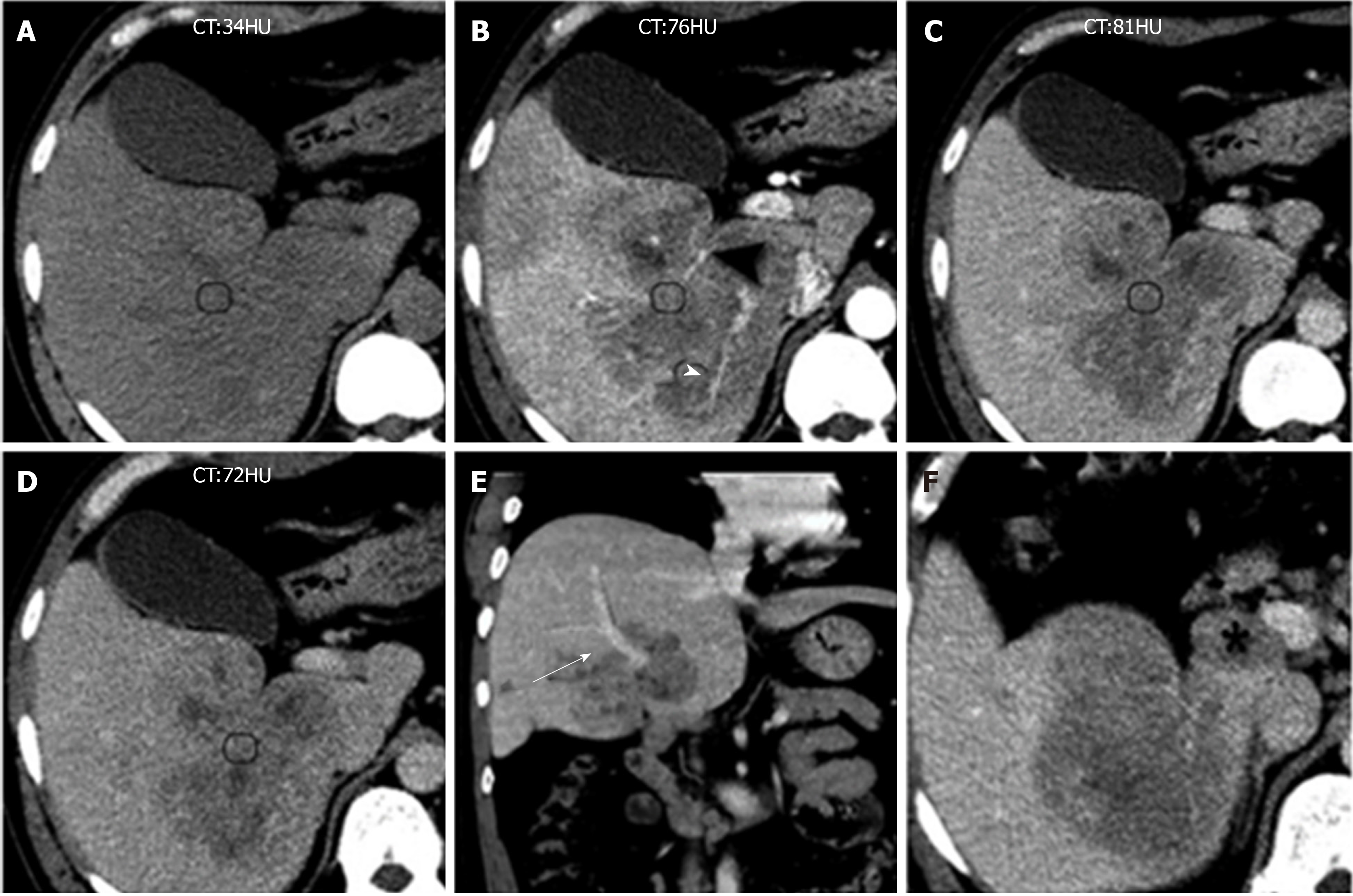
Figure 5 Computed tomography images of Case 2.
A: Unenhanced computed tomography (CT) image showing huge ill-defined heterogeneous hypodense masses; B-D: Multiphase enhanced CT images show a tumor with marked heterogeneous sustained hypoenhancement with internal necrosis. Engorged vascular structures (black arrowheads) were present in the mass; E: Coronal contrast-enhanced CT image showing the tumor near the portal vein (black arrow); and F: Axial contrast-enhanced CT image showing an enlarged lymph node seen in the hepatic hilar region (black star). Inflammatory pseudotumor-like follicular dendritic cell tumors from the right lobe of the liver were present in a 48-year-old Chinese man. CT: Computed tomography.
- Citation: Li HL, Liu HP, Guo GWJ, Chen ZH, Zhou FQ, Liu P, Liu JB, Wan R, Mao ZQ. Imaging findings of inflammatory pseudotumor-like follicular dendritic cell tumors of the liver: Two case reports and literature review. World J Gastroenterol 2019; 25(45): 6693-6703
- URL: https://www.wjgnet.com/1007-9327/full/v25/i45/6693.htm
- DOI: https://dx.doi.org/10.3748/wjg.v25.i45.6693


