Copyright
©The Author(s) 2019.
World J Gastroenterol. Jan 28, 2019; 25(4): 469-484
Published online Jan 28, 2019. doi: 10.3748/wjg.v25.i4.469
Published online Jan 28, 2019. doi: 10.3748/wjg.v25.i4.469
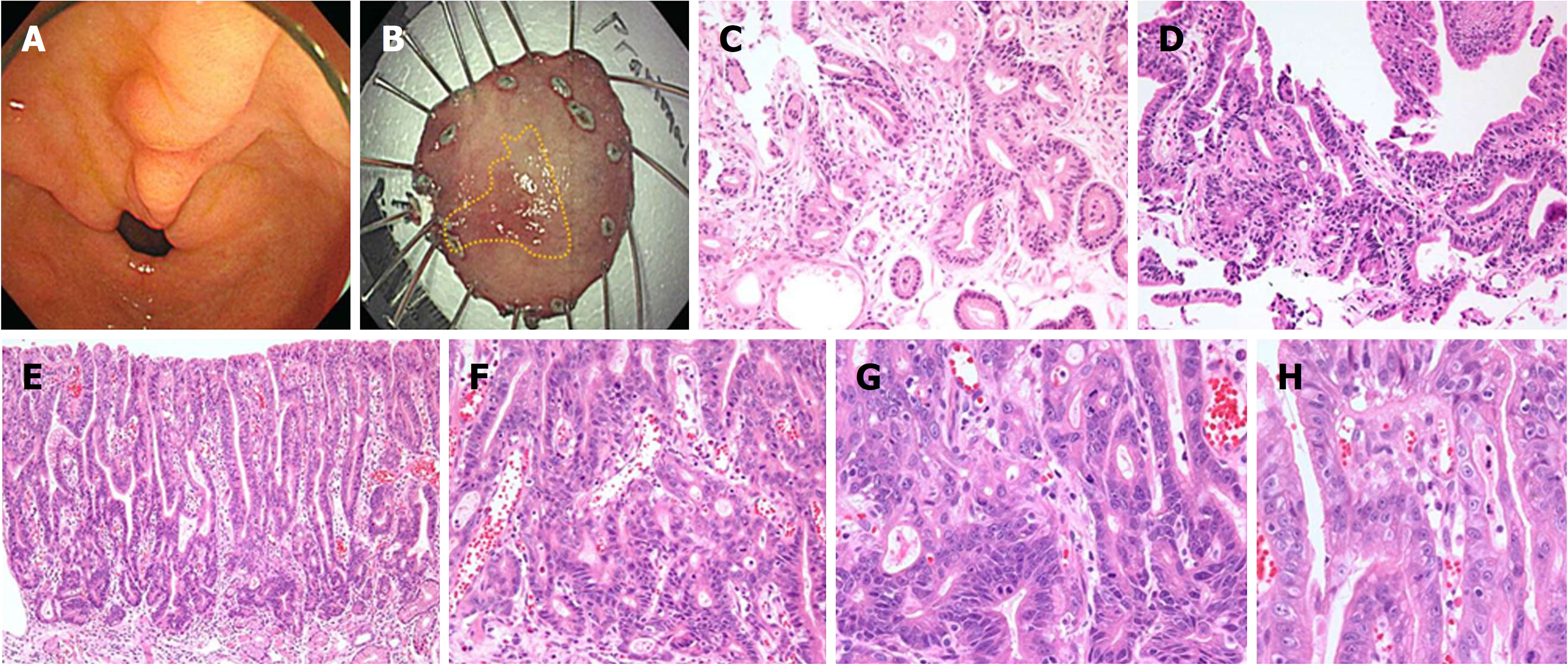
Figure 3 Representative images of Indefinite for neoplasm/dysplasia lesions with repeated diagnoses (from regenerating atypia to atypical epithelium), which were finally confirmed as well-differentiated adenocarcinoma at endoscopic submucosal dissection.
A: A lesion with mucosal irregularity and hyperemia is seen on the lesser curvature side of the prepylorus (indicated by arrows). B: After endoscopic submucosal dissection, the yellow line illustrates the boundary of the lesion confirmed in the pathology, measuring 1.7 cm × 1.0 cm. C: Regenerating atypia at the initial forceps biopsy shows focal glandular crowding with a basally-located, hyperchromatic nucleus. Glandular transition to the surrounding mucosa is observed. D: Atypical epithelium at follow-up biopsy after 598 d shows more crowded and tortuous glands. A few glands showed irregular distention. However, the hyperchromatic but basally-located nuclear atypia was mild. E: Endoscopic submucosal dissection was performed 2249 d later. It revealed well-differentiated adenocarcinoma. The low-power view shows surface maturation. F: The neoplastic tubules were prominent from the mid-portion of the tubular pit to the bottom of the gland; G: They form crowded, back-to-back, branched, and tortuous glands with disordered nuclei; H: Surface atypia is less prominent and mild nuclear atypia is observed. IFND: Indefinite for neoplasm/dysplasia.
- Citation: Kwon MJ, Kang HS, Kim HT, Choo JW, Lee BH, Hong SE, Park KH, Jung DM, Lim H, Soh JS, Moon SH, Kim JH, Park HR, Min SK, Seo JW, Choe JY. Treatment for gastric ‘indefinite for neoplasm/dysplasia’ lesions based on predictive factors. World J Gastroenterol 2019; 25(4): 469-484
- URL: https://www.wjgnet.com/1007-9327/full/v25/i4/469.htm
- DOI: https://dx.doi.org/10.3748/wjg.v25.i4.469


