Copyright
©The Author(s) 2019.
World J Gastroenterol. Jan 28, 2019; 25(4): 469-484
Published online Jan 28, 2019. doi: 10.3748/wjg.v25.i4.469
Published online Jan 28, 2019. doi: 10.3748/wjg.v25.i4.469
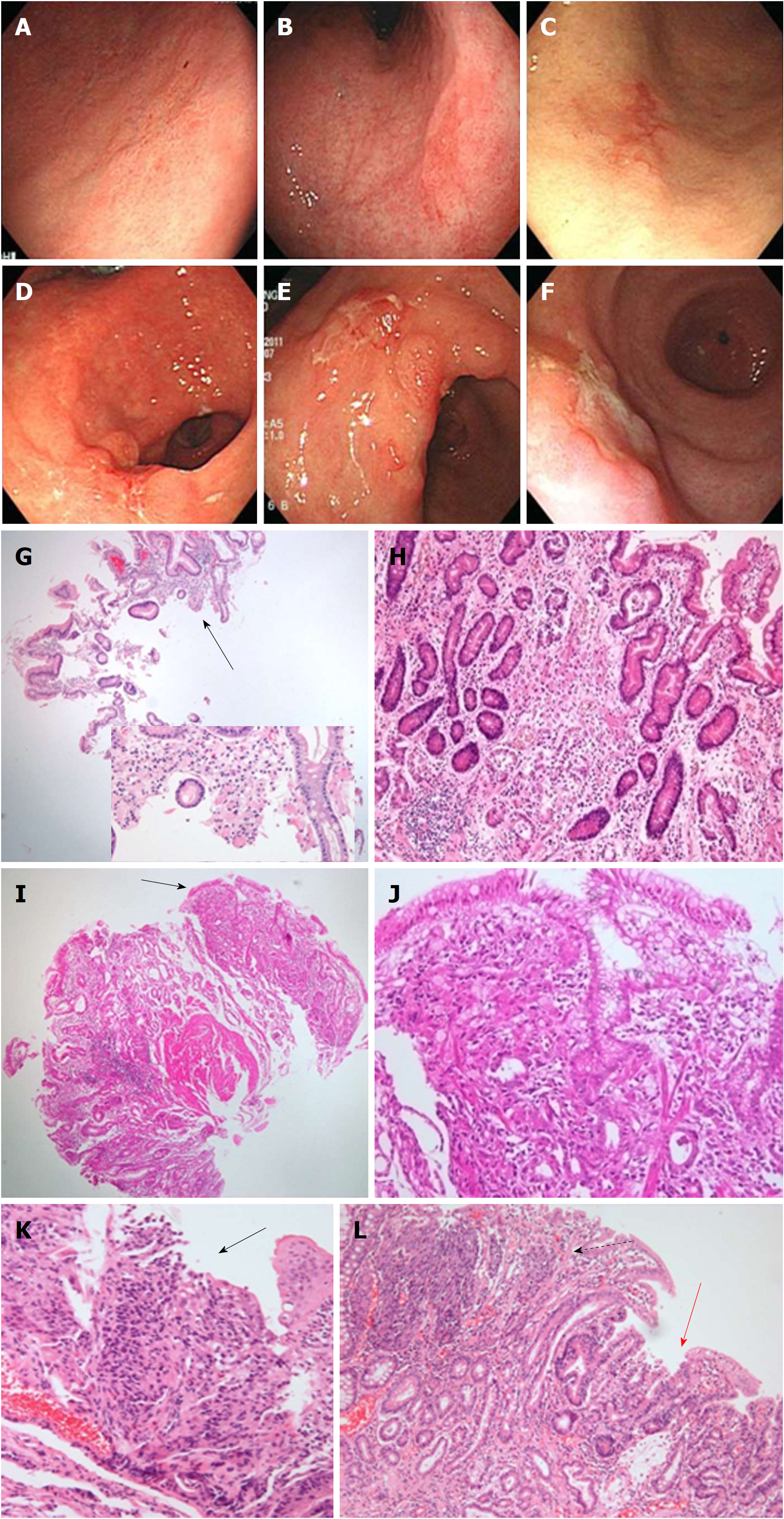
Figure 2 Representative cases of undifferentiated carcinoma.
Flat lesions with color change (A-C) and fold change with ulcerations (D-F) are shown at endoscopy. G: The case in panel A shows a few tumor cells (magnified in the inlet) in the endoscopic biopsy specimen (black arrow). H: The resected specimen shows poorly differentiated tubular adenocarcinoma with a very small tumor size (0.7 cm × 0.6 cm). I: The case in panel B shows a few tumor cells in the endoscopic biopsy specimen (black arrow). J: The resected specimen shows mixed signet ring cell carcinoma and poorly differentiated tubular adenocarcinoma with a very small tumor size (0.6 cm × 0.4 cm). K: The case in panel e shows a few tumor cells as squeezing artifact-like clusters in the endoscopic biopsy specimen (black arrow). L: The resected specimen shows mixed poorly differentiated (dotted arrow) and well-differentiated (red arrow) tubular adenocarcinoma.
- Citation: Kwon MJ, Kang HS, Kim HT, Choo JW, Lee BH, Hong SE, Park KH, Jung DM, Lim H, Soh JS, Moon SH, Kim JH, Park HR, Min SK, Seo JW, Choe JY. Treatment for gastric ‘indefinite for neoplasm/dysplasia’ lesions based on predictive factors. World J Gastroenterol 2019; 25(4): 469-484
- URL: https://www.wjgnet.com/1007-9327/full/v25/i4/469.htm
- DOI: https://dx.doi.org/10.3748/wjg.v25.i4.469


