Copyright
©The Author(s) 2019.
World J Gastroenterol. Sep 28, 2019; 25(36): 5569-5577
Published online Sep 28, 2019. doi: 10.3748/wjg.v25.i36.5569
Published online Sep 28, 2019. doi: 10.3748/wjg.v25.i36.5569
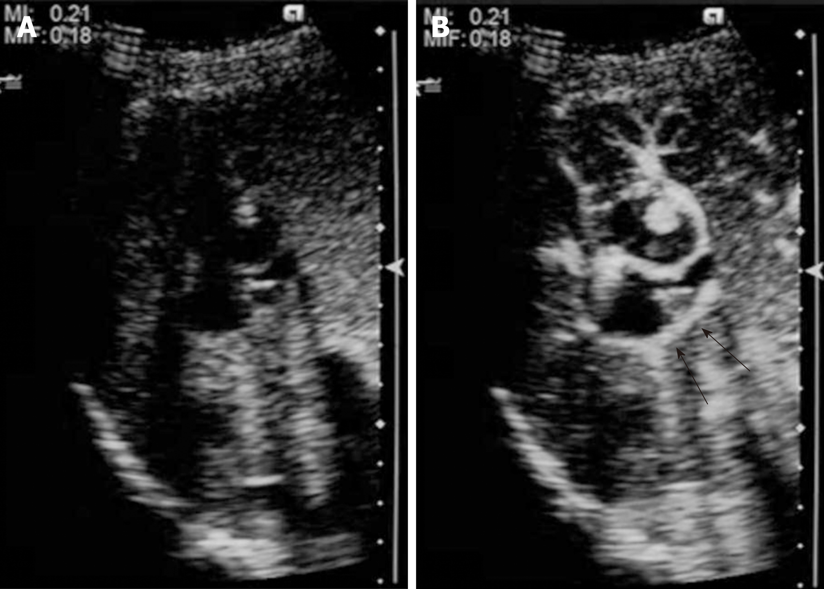
Figure 4 Ultrasonography performed 13 years after the initial clinical visit.
Contrast-enhanced ultrasonography shows a 7 mm hyperechoic papillary proliferation (A: Pre-enhancement; B: Arterial phase). Marginal hyperenhancement of the anterior tumor and early enhancement of the papillary proliferation were detected. Marginal enhancement of the posterior tumor (black arrows) was also detected, which suggests tumor spread to the posterior tumor.
- Citation: Hasebe T, Sawada K, Hayashi H, Nakajima S, Takahashi H, Hagiwara M, Imai K, Yuzawa S, Fujiya M, Furukawa H, Okumura T. Long-term growth of intrahepatic papillary neoplasms: A case report. World J Gastroenterol 2019; 25(36): 5569-5577
- URL: https://www.wjgnet.com/1007-9327/full/v25/i36/5569.htm
- DOI: https://dx.doi.org/10.3748/wjg.v25.i36.5569


