Copyright
©The Author(s) 2019.
World J Gastroenterol. Sep 28, 2019; 25(36): 5469-5482
Published online Sep 28, 2019. doi: 10.3748/wjg.v25.i36.5469
Published online Sep 28, 2019. doi: 10.3748/wjg.v25.i36.5469
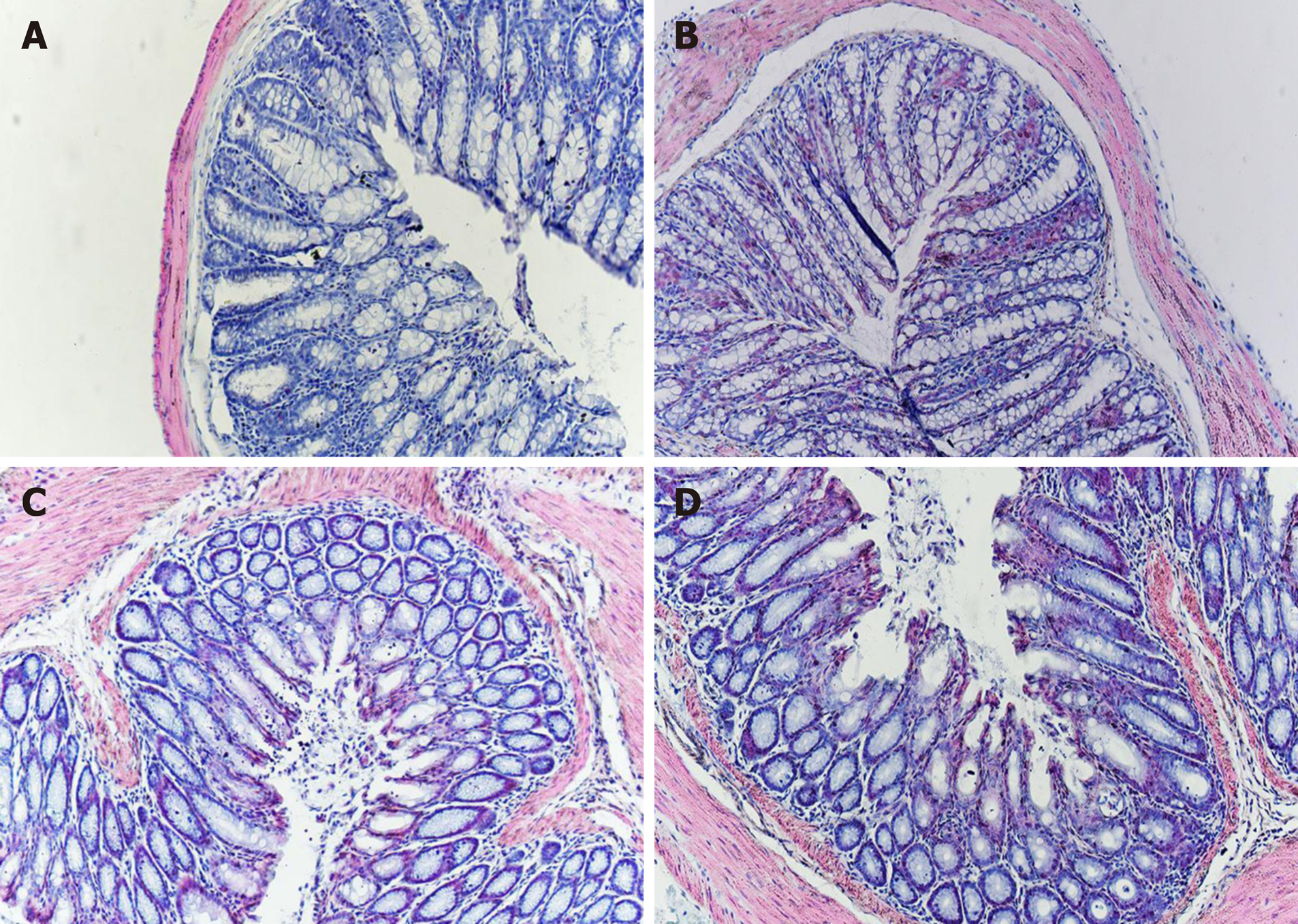
Figure 1 Photomicrographs of hematoxylin and eosin staining.
There was no difference with regard to morphology between control (A), IBS (B), IBS + C. butyricum (C), and IBS + NS (D) tissues in hematoxylin and eosin staining (original magnification, ×200) (n = 6 per group).
- Citation: Zhao Q, Yang WR, Wang XH, Li GQ, Xu LQ, Cui X, Liu Y, Zuo XL. Clostridium butyricum alleviates intestinal low-grade inflammation in TNBS-induced irritable bowel syndrome in mice by regulating functional status of lamina propria dendritic cells. World J Gastroenterol 2019; 25(36): 5469-5482
- URL: https://www.wjgnet.com/1007-9327/full/v25/i36/5469.htm
- DOI: https://dx.doi.org/10.3748/wjg.v25.i36.5469


