Copyright
©The Author(s) 2019.
World J Gastroenterol. Sep 7, 2019; 25(33): 4892-4903
Published online Sep 7, 2019. doi: 10.3748/wjg.v25.i33.4892
Published online Sep 7, 2019. doi: 10.3748/wjg.v25.i33.4892
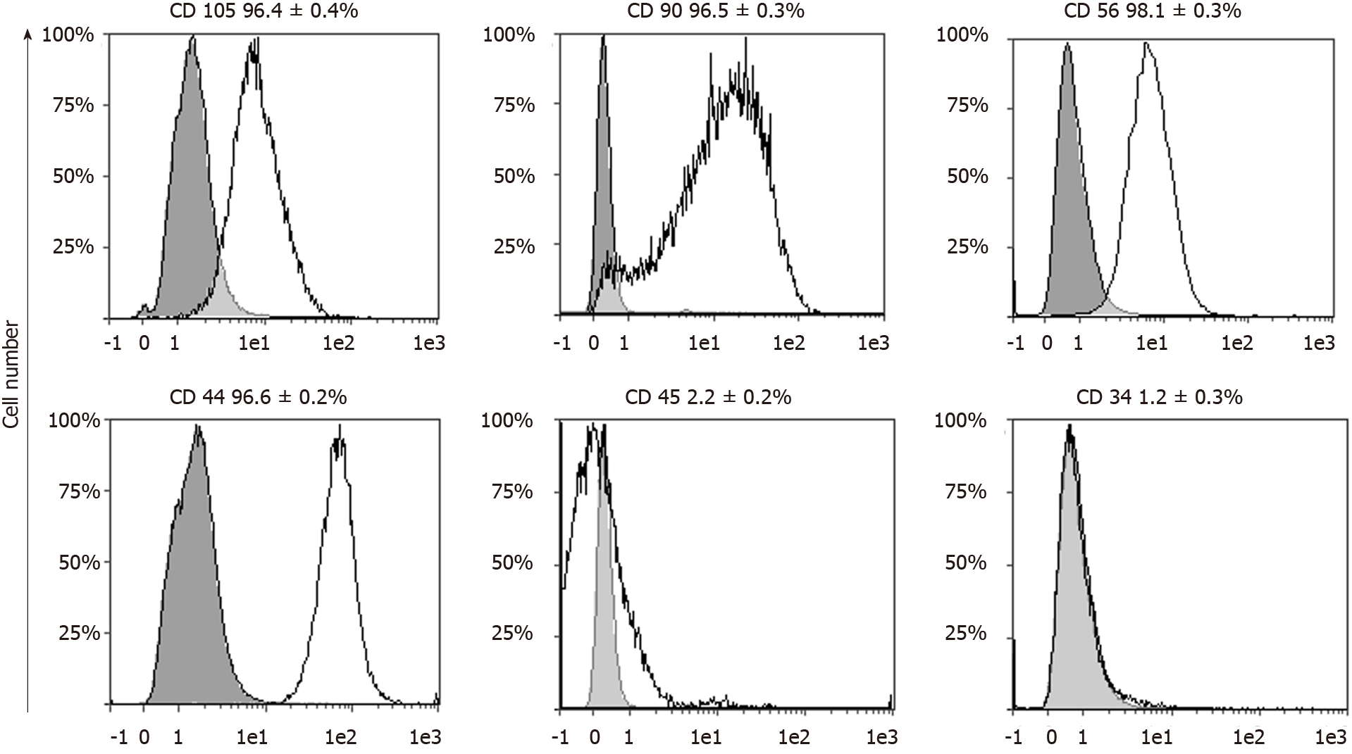
Figure 3 Flowcytometric analysis of cell-surface markers in porcine vascular wall mesenchymal stromal cells.
Each graph shows the percentage of cells expressing the specific marker reported [white area under the curve (AUC)] and the relative negative control (gray AUC, cells not incubated with any antibodies). This analysis confirmed the mesenchymal stromal cell-like immune profile of porcine vascular wall mesenchymal stromal cells: CD105, CD90, CD56, CD44 were highly expressed (> 96%) while the hematopoietic markers CD45 and CD43 were nearly absent (< 2.5%). AUC: Area under the curve.
- Citation: Dothel G, Bernardini C, Zannoni A, Spirito MR, Salaroli R, Bacci ML, Forni M, Ponti FD. Ex vivo effect of vascular wall stromal cells secretome on enteric ganglia. World J Gastroenterol 2019; 25(33): 4892-4903
- URL: https://www.wjgnet.com/1007-9327/full/v25/i33/4892.htm
- DOI: https://dx.doi.org/10.3748/wjg.v25.i33.4892


