Copyright
©The Author(s) 2019.
World J Gastroenterol. Sep 7, 2019; 25(33): 4892-4903
Published online Sep 7, 2019. doi: 10.3748/wjg.v25.i33.4892
Published online Sep 7, 2019. doi: 10.3748/wjg.v25.i33.4892
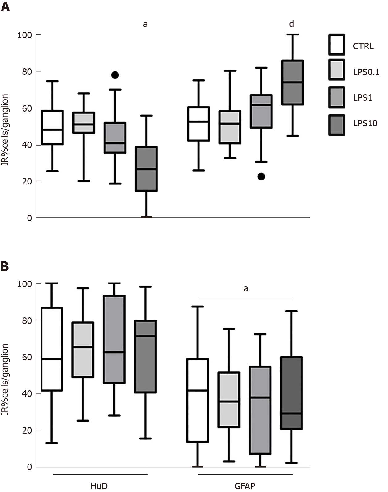
Figure 2 Effect of increasing concentration of lipopolysaccharide on enteric ganglia’ HUD+ neurons and GFAP+ glial cells.
A: In guinea pig-derived enteric ganglia - lipopolysaccharide (LPS) at 10 µg/mL decreased number of HuD-immunoreactive (HuD-IR) neurons (left columns) and increased proliferation of glial fibrillary acidic protein-immunoreactive (GFAP-IR) glial cells (right columns - HuD-IR neurons LPS10 vs CTRL, 22.3%, aP < 0.05; GFAP-IR glial cells LPS10 vs CTRL, +22.2%, dP < 0.01); B: Conversely, in pig enteric ganglia the number of glial cells at every LPS concentration tested did not change and was significatively lower compared to ganglionic neurons. aP < 0.05. LPS: lipopolysaccharide; GFAP-IR: Glial fibrillary acidic protein-immunoreactive; HuD-IR: HuD-immunoreactive.
- Citation: Dothel G, Bernardini C, Zannoni A, Spirito MR, Salaroli R, Bacci ML, Forni M, Ponti FD. Ex vivo effect of vascular wall stromal cells secretome on enteric ganglia. World J Gastroenterol 2019; 25(33): 4892-4903
- URL: https://www.wjgnet.com/1007-9327/full/v25/i33/4892.htm
- DOI: https://dx.doi.org/10.3748/wjg.v25.i33.4892


