Copyright
©The Author(s) 2019.
World J Gastroenterol. Aug 28, 2019; 25(32): 4682-4695
Published online Aug 28, 2019. doi: 10.3748/wjg.v25.i32.4682
Published online Aug 28, 2019. doi: 10.3748/wjg.v25.i32.4682
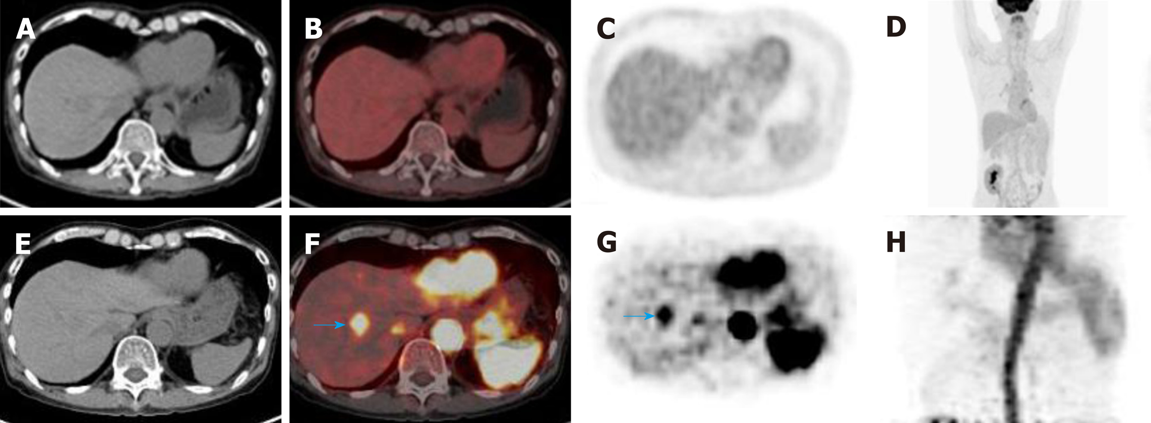
Figure 3 Early dynamic 2-deoxy-2-(18F)fluoro-D-glucose positron-emission tomography-computed tomography detected a tumor that was missed on conventional 2-deoxy-2-(18F)fluoro-D-glucose positron-emission tomography-computed tomography in a 64-year-old patient with hepatocellular carcinoma.
A-D: 2-deoxy-2-(18F)fluoro-D-glucose positron-emission tomography-computed tomography (18F-FDG PET-CT) showed that there was no increased 18F-FDG uptake in the lesion on conventional 18F-FDG PET-CT; E-H: Early dynamic 18F-FDG PET-CT showed focal 18F-FDG hyperperfusion in the upper segment of the anterior lobe of the liver, and the size of the lesion was 1.7 × 1.9 cm (blue arrow in F and G). 18F-FDG: 2-deoxy-2-(18F)fluoro-D-glucose; CT: Computed tomography; PET: Positron-emission tomography.
- Citation: Lu RC, She B, Gao WT, Ji YH, Xu DD, Wang QS, Wang SB. Positron-emission tomography for hepatocellular carcinoma: Current status and future prospects. World J Gastroenterol 2019; 25(32): 4682-4695
- URL: https://www.wjgnet.com/1007-9327/full/v25/i32/4682.htm
- DOI: https://dx.doi.org/10.3748/wjg.v25.i32.4682


