Copyright
©The Author(s) 2019.
World J Gastroenterol. Aug 28, 2019; 25(32): 4682-4695
Published online Aug 28, 2019. doi: 10.3748/wjg.v25.i32.4682
Published online Aug 28, 2019. doi: 10.3748/wjg.v25.i32.4682
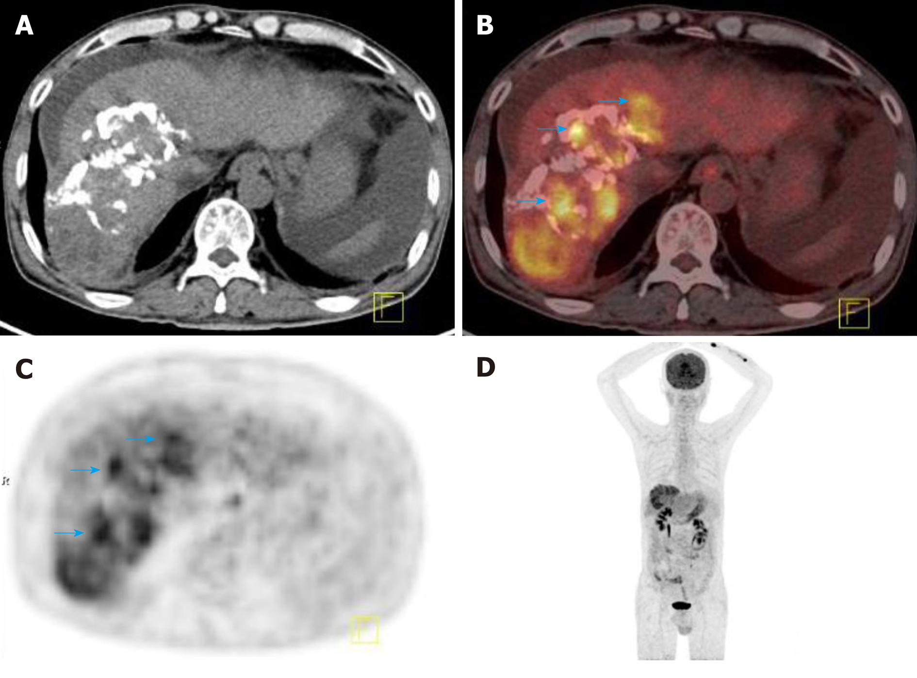
Figure 1 2-Deoxy-2-(18F)fluoro-D-glucose positron-emission tomography-computed tomography detected tumor recurrence after intervention therapy in a 58-year-old male patient with hepatocellular carcinoma.
A: Cross-sectional computed tomography (CT) image showing a large sheet of lipiodol deposition in the right lobe of live after HCC intervention therapy; B: Cross-sectional positron-emission tomography (PET-CT) fusion image showing increased 18F-FDG uptake in and around the area of lipiodol deposition (blue arrow); the size of the lesion was 5.8 × 13.3 cm; C: Cross-sectional PET image showing increased 18F-FDG uptake in the right lobe of the liver; D: Maximum intensity projection image showing increased 18F-FDG uptake in the right lobe of the liver. 18F-FDG: 2-deoxy-2-(18F)fluoro-D-glucose; CT: Computed tomography; PET: Positron-emission tomography.
- Citation: Lu RC, She B, Gao WT, Ji YH, Xu DD, Wang QS, Wang SB. Positron-emission tomography for hepatocellular carcinoma: Current status and future prospects. World J Gastroenterol 2019; 25(32): 4682-4695
- URL: https://www.wjgnet.com/1007-9327/full/v25/i32/4682.htm
- DOI: https://dx.doi.org/10.3748/wjg.v25.i32.4682


