Copyright
©The Author(s) 2019.
World J Gastroenterol. Aug 21, 2019; 25(31): 4320-4342
Published online Aug 21, 2019. doi: 10.3748/wjg.v25.i31.4320
Published online Aug 21, 2019. doi: 10.3748/wjg.v25.i31.4320
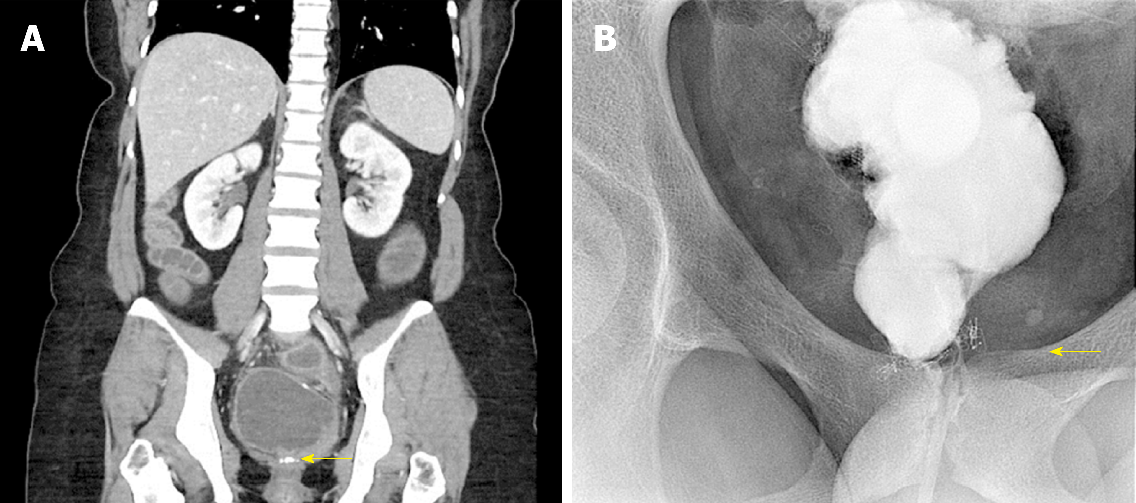
Figure 5 Obstruction to pouch outflow usually occurs at the level of the anastomosis.
A: A coronal computed tomography image demonstrating a pouch outlet stricture. A stricture at the level of the anastomosis (arrow) caused a dilated pouch that could not empty without intubation; B: A pouchogram of the same patient confirmed an anastomotic stricture that eventually yielded to serial Hegar dilations.
- Citation: Ng KS, Gonsalves SJ, Sagar PM. Ileal-anal pouches: A review of its history, indications, and complications. World J Gastroenterol 2019; 25(31): 4320-4342
- URL: https://www.wjgnet.com/1007-9327/full/v25/i31/4320.htm
- DOI: https://dx.doi.org/10.3748/wjg.v25.i31.4320


