Copyright
©The Author(s) 2019.
World J Gastroenterol. Aug 14, 2019; 25(30): 4213-4221
Published online Aug 14, 2019. doi: 10.3748/wjg.v25.i30.4213
Published online Aug 14, 2019. doi: 10.3748/wjg.v25.i30.4213
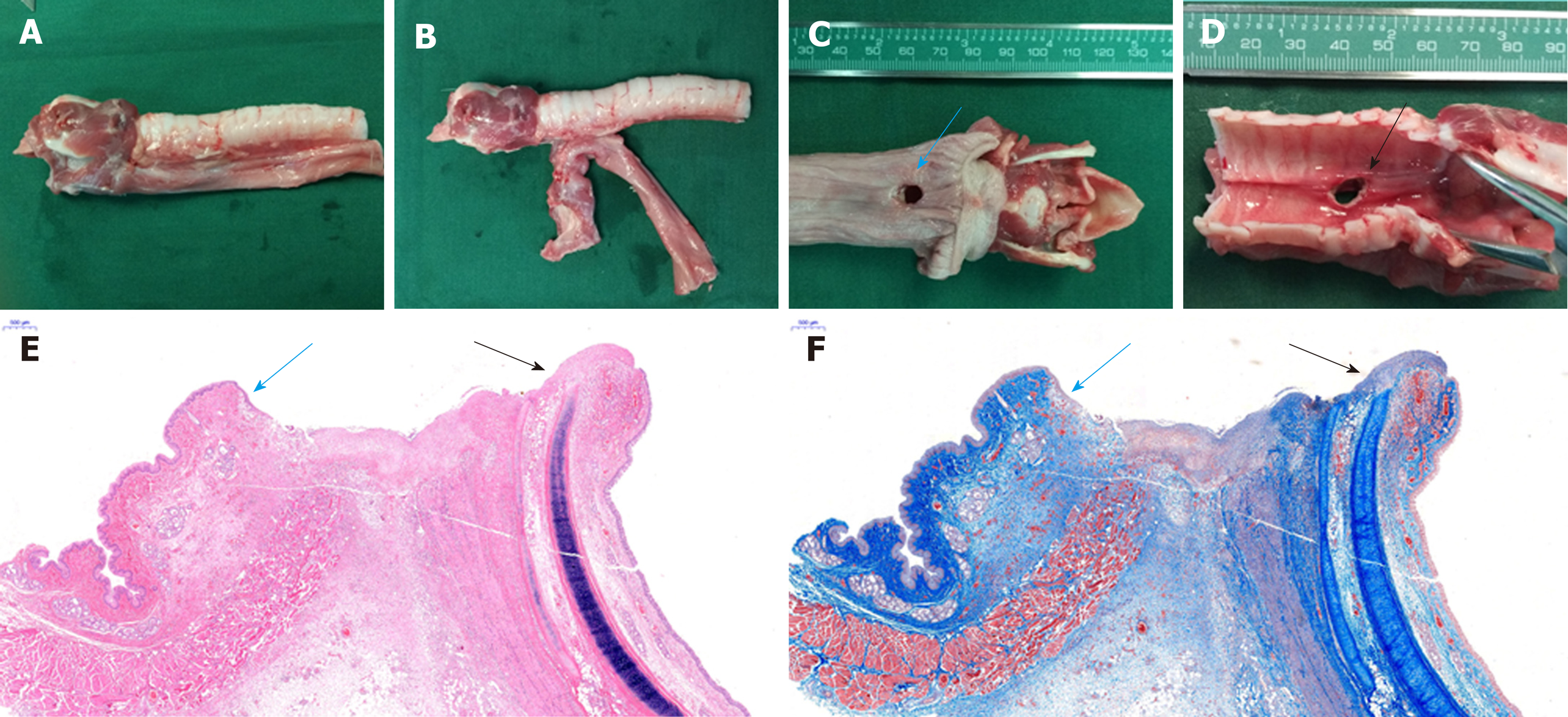
Figure 5 Gross and histological observations.
A: Gross specimens of the trachea and esophagus; B: There was no tissue adhesion between the trachea and the esophagus, except around the fistula; C: Gross specimen showing the fistula in the esophagus; D: Gross specimen showing the fistula in the trachea; E and F: Histological analysis demonstrated that the esophageal mucosa (blue arrow) and pseudostratified ciliated columnar epithelium (black arrow) were absent at the site of the fistula.
- Citation: Gao Y, Wu RQ, Lv Y, Yan XP. Novel magnetic compression technique for establishment of a canine model of tracheoesophageal fistula. World J Gastroenterol 2019; 25(30): 4213-4221
- URL: https://www.wjgnet.com/1007-9327/full/v25/i30/4213.htm
- DOI: https://dx.doi.org/10.3748/wjg.v25.i30.4213


