Copyright
©The Author(s) 2019.
World J Gastroenterol. Aug 14, 2019; 25(30): 4213-4221
Published online Aug 14, 2019. doi: 10.3748/wjg.v25.i30.4213
Published online Aug 14, 2019. doi: 10.3748/wjg.v25.i30.4213
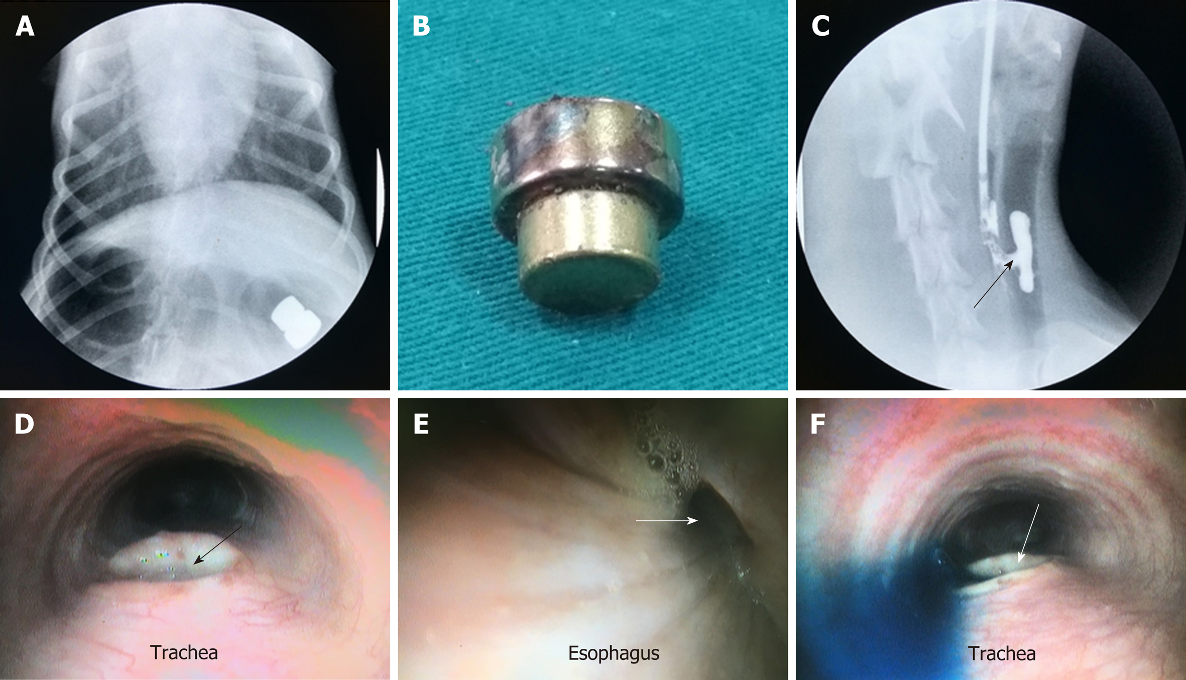
Figure 4 Results after implantation.
A: 6 d after magnet implantation: X-ray showing that the magnets were located in the digestive tract; B: 8 d after magnet implantation: the magnets were expelled from the animal through the digestive tract; C: 8 d after magnet implantation: esophageal angiography showing the contrast media flowing from the esophagus into the trachea; D: Bronchoscopy showing a fistula located in the posterior wall of the trachea; E: Gastroscopy showing a fistula located in the anterior wall of the esophagus; F: Gastroscopy/bronchoscopy showing methylene blue flowing from the esophagus into the trachea.
- Citation: Gao Y, Wu RQ, Lv Y, Yan XP. Novel magnetic compression technique for establishment of a canine model of tracheoesophageal fistula. World J Gastroenterol 2019; 25(30): 4213-4221
- URL: https://www.wjgnet.com/1007-9327/full/v25/i30/4213.htm
- DOI: https://dx.doi.org/10.3748/wjg.v25.i30.4213


