Copyright
©The Author(s) 2019.
World J Gastroenterol. Jan 21, 2019; 25(3): 367-377
Published online Jan 21, 2019. doi: 10.3748/wjg.v25.i3.367
Published online Jan 21, 2019. doi: 10.3748/wjg.v25.i3.367
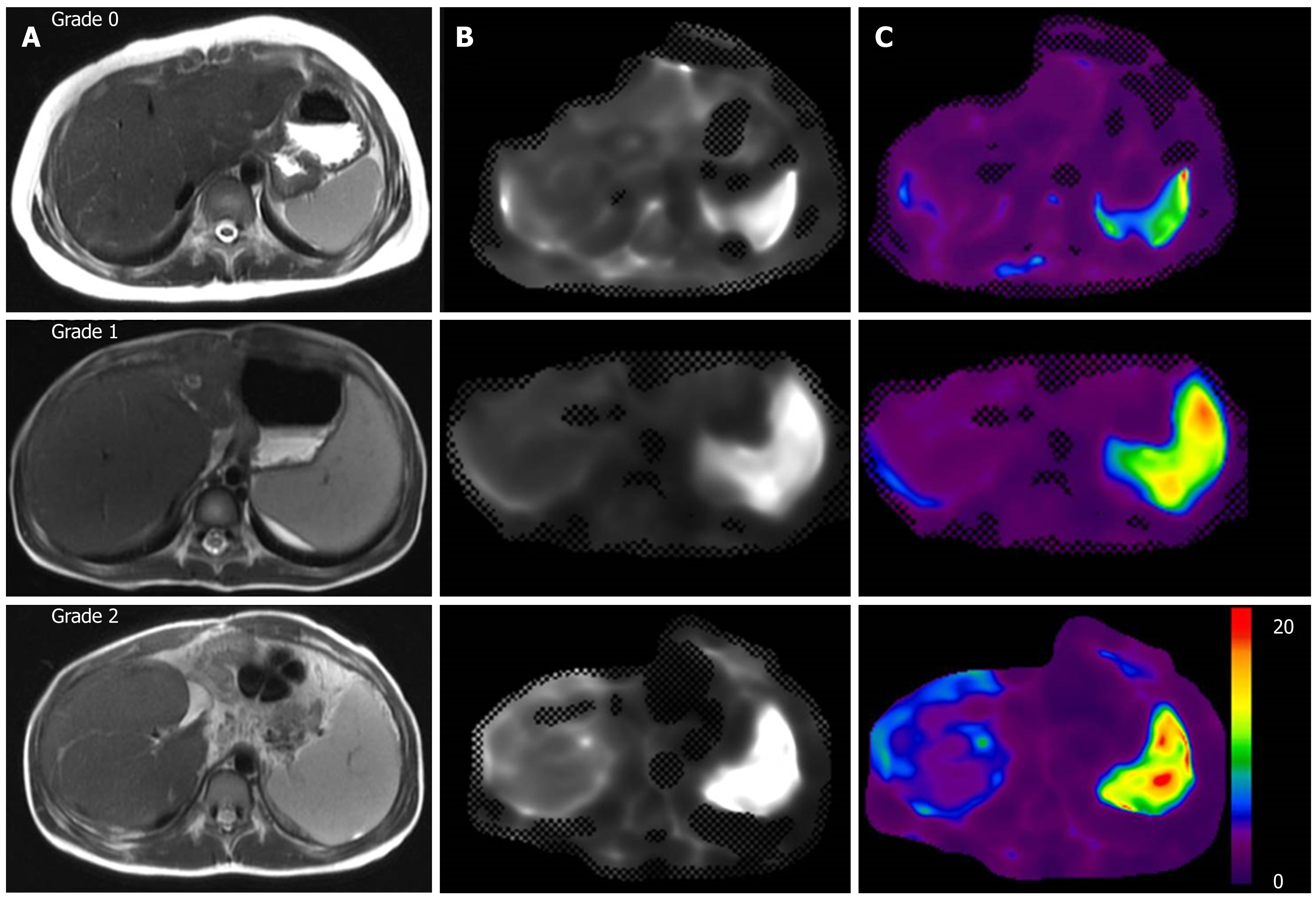
Figure 2 Liver magnetic resonance imaging of biliary atresia patients with different varices grades.
Liver magnetic resonance imaging images are shown from three respective patients with grade 0 (first row), grade 1 (second row) and grade 2 (third row) gastroesophageal varices, including (A) axial single-shot fast spin-echo T2-weighted images, (B) magnitude images and (C) post-processed shear stiffness maps with color-coded elastograms from 0 to 20 kPa. The elastograms display the progressively increasing splenic stiffness in these patients.
- Citation: Yoon H, Shin HJ, Kim MJ, Han SJ, Koh H, Kim S, Lee MJ. Predicting gastroesophageal varices through spleen magnetic resonance elastography in pediatric liver fibrosis. World J Gastroenterol 2019; 25(3): 367-377
- URL: https://www.wjgnet.com/1007-9327/full/v25/i3/367.htm
- DOI: https://dx.doi.org/10.3748/wjg.v25.i3.367


