Copyright
©The Author(s) 2019.
World J Gastroenterol. Jan 21, 2019; 25(3): 300-307
Published online Jan 21, 2019. doi: 10.3748/wjg.v25.i3.300
Published online Jan 21, 2019. doi: 10.3748/wjg.v25.i3.300
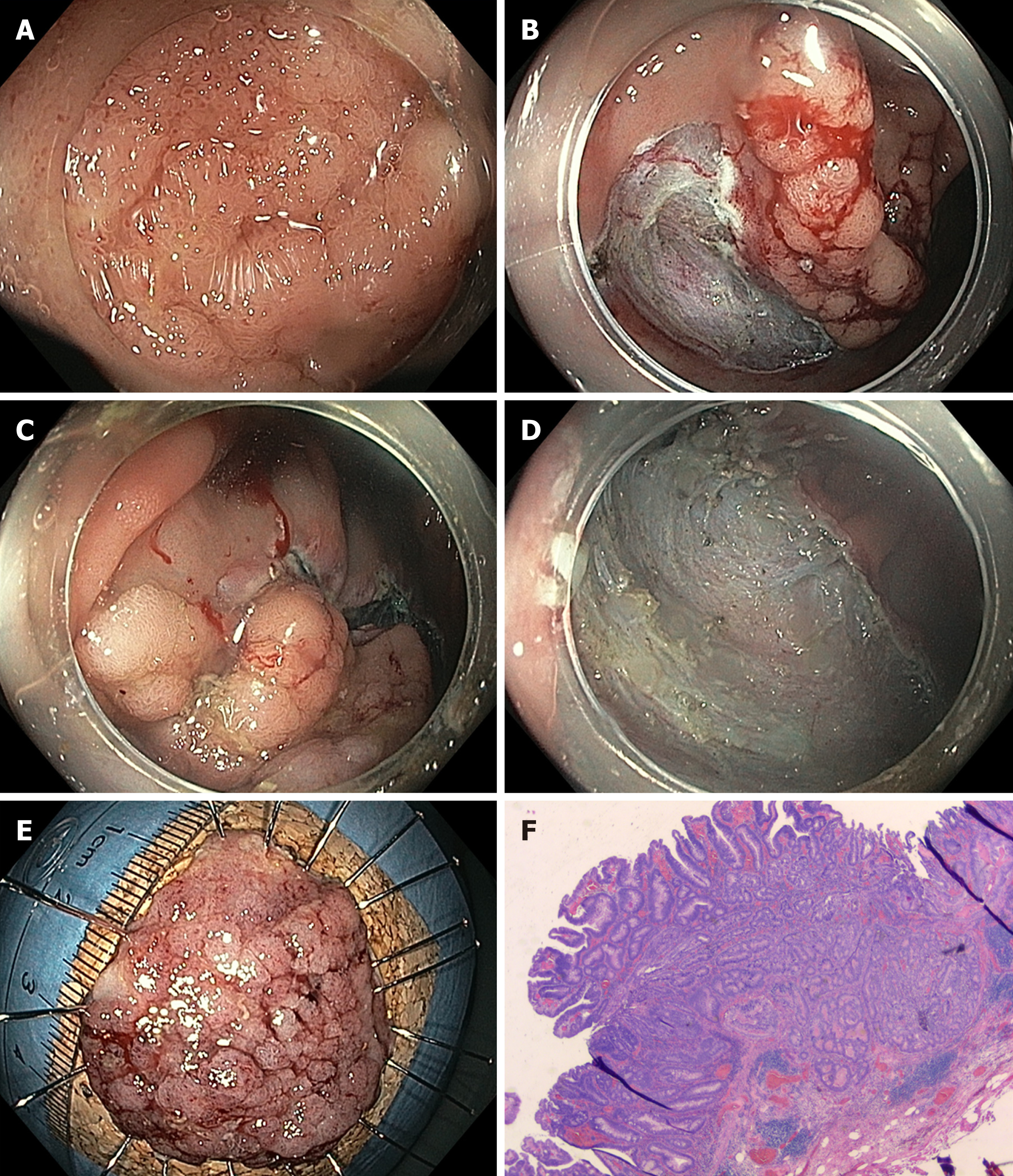
Figure 2 Endoscopic submucosal dissection.
A: Endoscopic aspect of a large sessile lesion (Paris 0-Is/0-IIa; lateral spreading tumor, granular type) in the cecum. B: Start of endoscopic submucosal dissection at the proximal site. C: Mucosal incision at the distal margin. D: Completed resection with resection bed in the cecum. E: Resected specimen on corkboard. F: Histopathology: tubulovillous adenoma with focal high-grade intraepithelial neoplasia (hematoxylin/eosine, magnification: 80-fold).
- Citation: Dumoulin FL, Hildenbrand R. Endoscopic resection techniques for colorectal neoplasia: Current developments. World J Gastroenterol 2019; 25(3): 300-307
- URL: https://www.wjgnet.com/1007-9327/full/v25/i3/300.htm
- DOI: https://dx.doi.org/10.3748/wjg.v25.i3.300


