Copyright
©The Author(s) 2019.
World J Gastroenterol. Aug 7, 2019; 25(29): 3941-3955
Published online Aug 7, 2019. doi: 10.3748/wjg.v25.i29.3941
Published online Aug 7, 2019. doi: 10.3748/wjg.v25.i29.3941
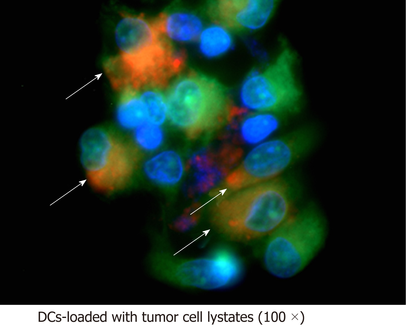
Figure 3 Investigation of tumour antigen on dendritic cells by fluorescence microscopy.
Dendritic cells were stained with CellTracker™ Green CMFDA before loading with honokiol-derived tumour cell lysates pre-stained with CellTracker™ Red CMPTX. Antigen load was verified by visualisation of tumour antigen (red) within dendritic cells (green). Arrowheads indicate co-expression of green and red fluorescence. DCs: Dendritic cells.
- Citation: Jiraviriyakul A, Songjang W, Kaewthet P, Tanawatkitichai P, Bayan P, Pongcharoen S. Honokiol-enhanced cytotoxic T lymphocyte activity against cholangiocarcinoma cells mediated by dendritic cells pulsed with damage-associated molecular patterns. World J Gastroenterol 2019; 25(29): 3941-3955
- URL: https://www.wjgnet.com/1007-9327/full/v25/i29/3941.htm
- DOI: https://dx.doi.org/10.3748/wjg.v25.i29.3941


