Copyright
©The Author(s) 2019.
World J Gastroenterol. Jul 21, 2019; 25(27): 3619-3633
Published online Jul 21, 2019. doi: 10.3748/wjg.v25.i27.3619
Published online Jul 21, 2019. doi: 10.3748/wjg.v25.i27.3619
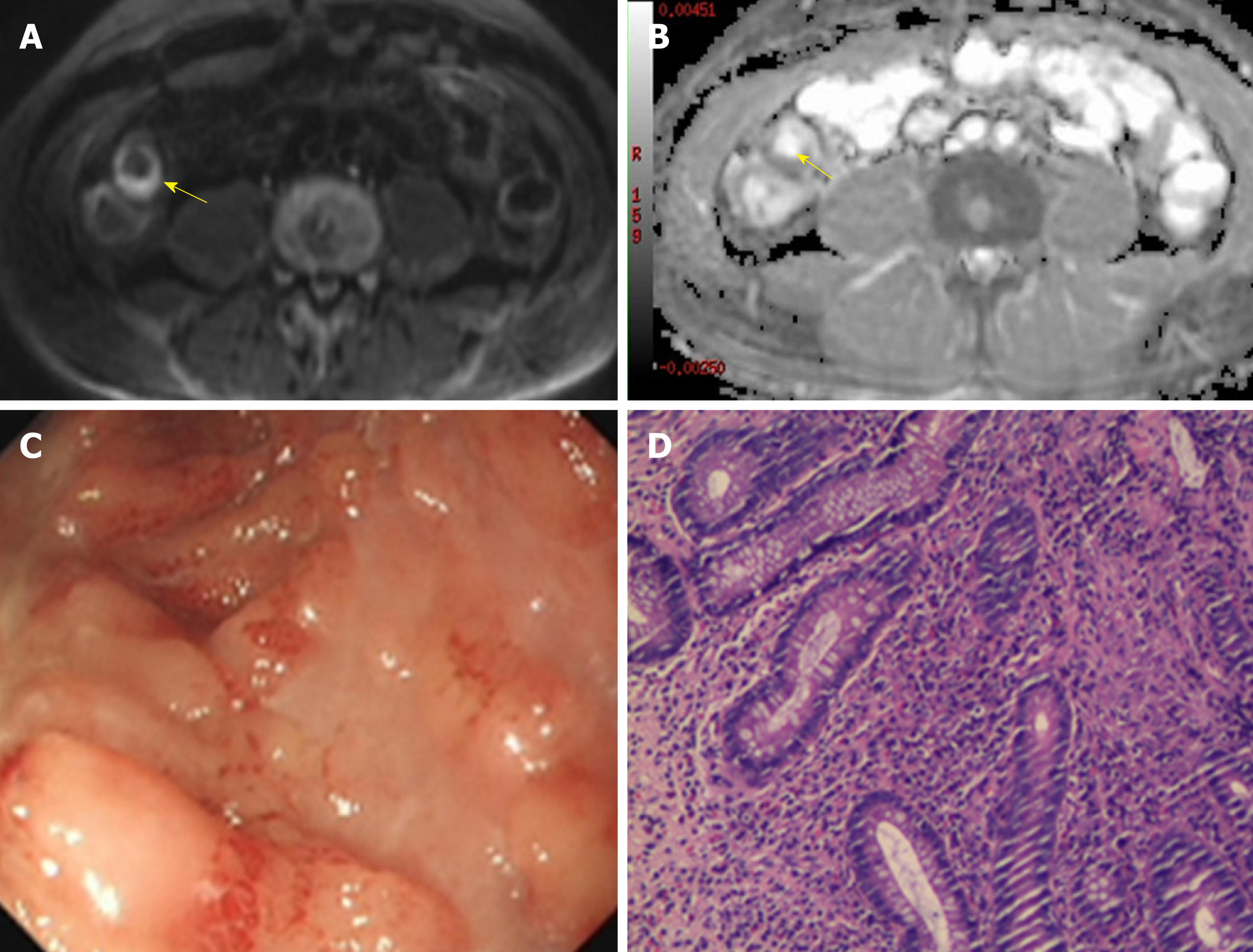
Figure 2 A 48-year-old man with Crohn’s disease involving the terminal ileum.
A: Axial diffusion-weighted imaging with b = 800 s/mm2 demonstrated high signal intensity of the terminal ileum (arrow). B: The terminal ileum was hypointense (arrow) on the corresponding apparent diffusion coefficient map. C: Ileocolonoscopy showed mucosal ulcers. D: Histopathology demonstrated neutrophil infiltration.
- Citation: Yu H, Shen YQ, Tan FQ, Zhou ZL, Li Z, Hu DY, Morelli JN. Quantitative diffusion-weighted magnetic resonance enterography in ileal Crohn's disease: A systematic analysis of intra and interobserver reproducibility. World J Gastroenterol 2019; 25(27): 3619-3633
- URL: https://www.wjgnet.com/1007-9327/full/v25/i27/3619.htm
- DOI: https://dx.doi.org/10.3748/wjg.v25.i27.3619


