Copyright
©The Author(s) 2019.
World J Gastroenterol. Jul 7, 2019; 25(25): 3207-3217
Published online Jul 7, 2019. doi: 10.3748/wjg.v25.i25.3207
Published online Jul 7, 2019. doi: 10.3748/wjg.v25.i25.3207
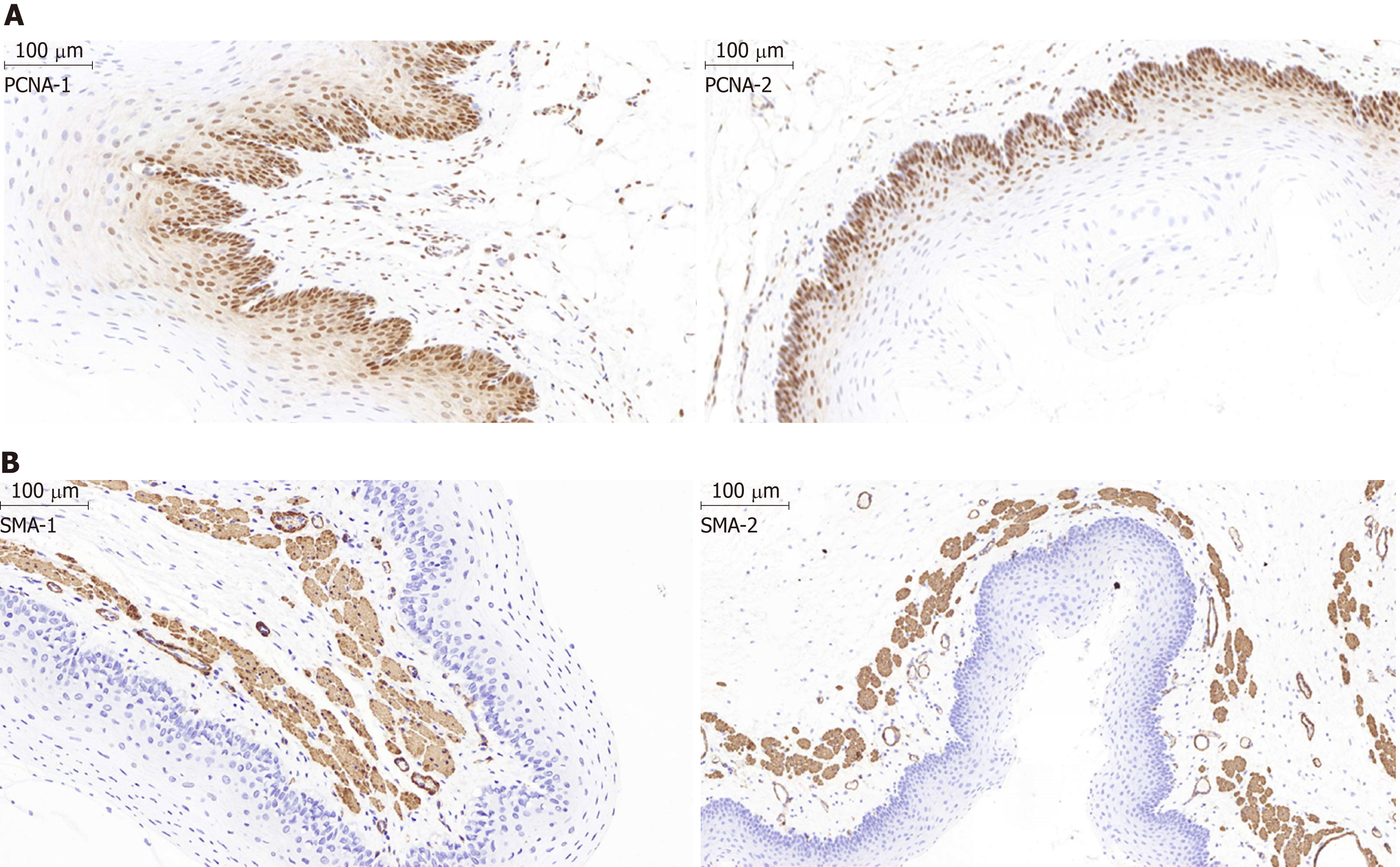
Figure 6 Histological examination of the epithelial layer and the α-smooth muscle layer of the esophagus after stent insertion.
A: Representative images show the proliferating cell nuclear antigen (PCNA) staining in the epithelial layer of the esophagus from the stent group and the normal control group. The percentage of PCNA-positive proliferating cells in the epithelial layer in the control group (PCNA-1) was similar to that in the magnesium-silicone stent group (PCNA-2) (magnification, ×100). B: Representative images showing the α-smooth muscle (SMA) staining. The thickness of the epithelial and SMA-positive layers did not differ between the normal control group (SMA-1) and magnesium-silicone stent group (SMA-2) (magnification, ×200).
- Citation: Yang K, Cao J, Yuan TW, Zhu YQ, Zhou B, Cheng YS. Silicone-covered biodegradable magnesium stent for treating benign esophageal stricture in a rabbit model. World J Gastroenterol 2019; 25(25): 3207-3217
- URL: https://www.wjgnet.com/1007-9327/full/v25/i25/3207.htm
- DOI: https://dx.doi.org/10.3748/wjg.v25.i25.3207


