Copyright
©The Author(s) 2019.
World J Gastroenterol. Jul 7, 2019; 25(25): 3108-3115
Published online Jul 7, 2019. doi: 10.3748/wjg.v25.i25.3108
Published online Jul 7, 2019. doi: 10.3748/wjg.v25.i25.3108
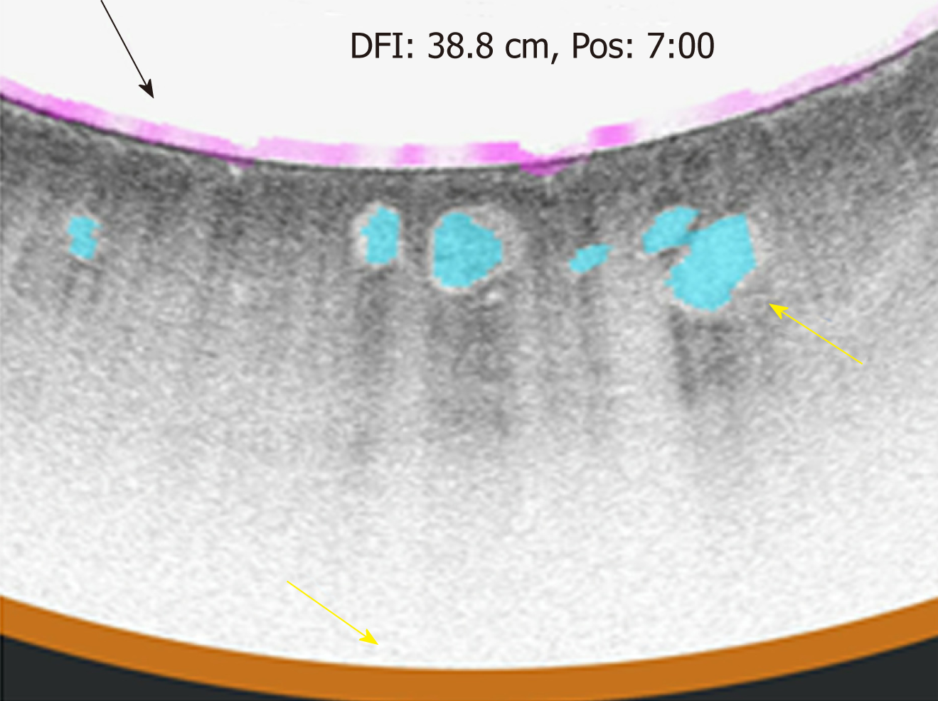
Figure 4 Volumetric laser endomicroscopy with artificial intelligence showing an up close snap shot of the abnormal area of overlap between three features of dysplasia (orange is lack of layering, blue is glandular structures, and pink is a hyper-reflective surface).
- Citation: Cerrone SA, Trindade AJ. Advanced imaging in surveillance of Barrett’s esophagus: Is the juice worth the squeeze? World J Gastroenterol 2019; 25(25): 3108-3115
- URL: https://www.wjgnet.com/1007-9327/full/v25/i25/3108.htm
- DOI: https://dx.doi.org/10.3748/wjg.v25.i25.3108


