Copyright
©The Author(s) 2019.
World J Gastroenterol. Jun 28, 2019; 25(24): 3091-3107
Published online Jun 28, 2019. doi: 10.3748/wjg.v25.i24.3091
Published online Jun 28, 2019. doi: 10.3748/wjg.v25.i24.3091
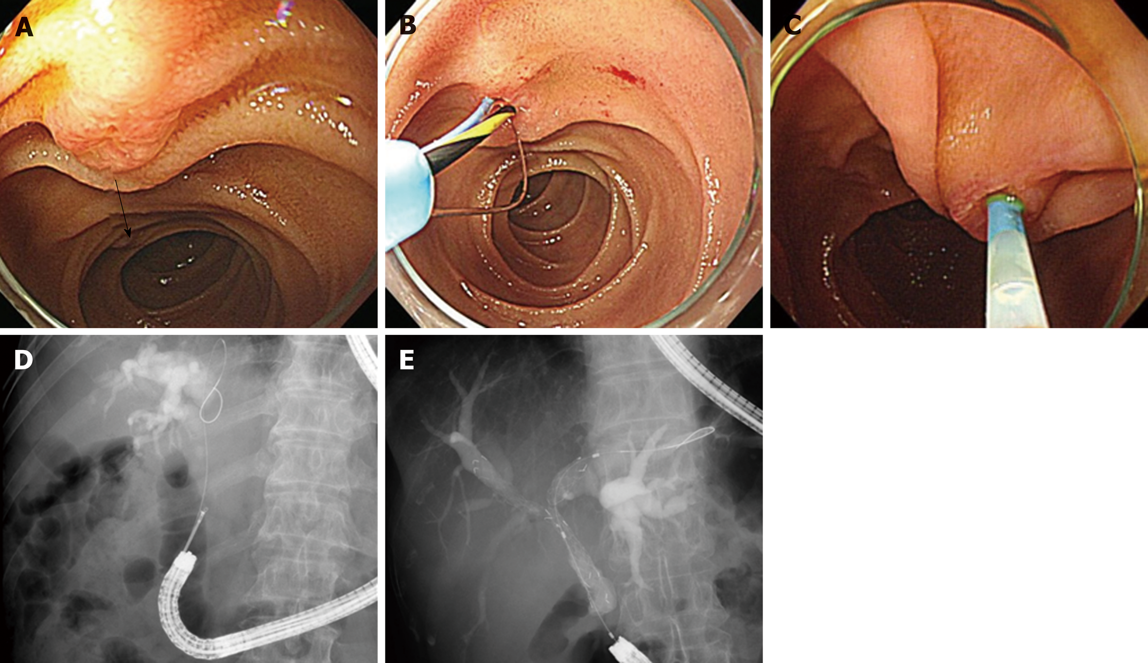
Figure 3 Cap-fitting forward-viewing endoscopy.
A: Naïve papilla; It is difficult to obtain en face view. The direction of bile duct is reversed (arrow); B: Gastroscope of 7 o’clock position working channel; Sphincter management with inverted sphincterotome; C: Pediatric colonoscope of 5 o’clock position working channel; D: Endobiliary biopsy was performed in distal common bile duct stricture; E: Bilateral uncovered metal stents were inserted in the malignant hilar stricture.
- Citation: Park TY, Song TJ. Recent advances in endoscopic retrograde cholangiopancreatography in Billroth II gastrectomy patients: A systematic review. World J Gastroenterol 2019; 25(24): 3091-3107
- URL: https://www.wjgnet.com/1007-9327/full/v25/i24/3091.htm
- DOI: https://dx.doi.org/10.3748/wjg.v25.i24.3091


