Copyright
©The Author(s) 2019.
World J Gastroenterol. Jun 28, 2019; 25(24): 3030-3043
Published online Jun 28, 2019. doi: 10.3748/wjg.v25.i24.3030
Published online Jun 28, 2019. doi: 10.3748/wjg.v25.i24.3030
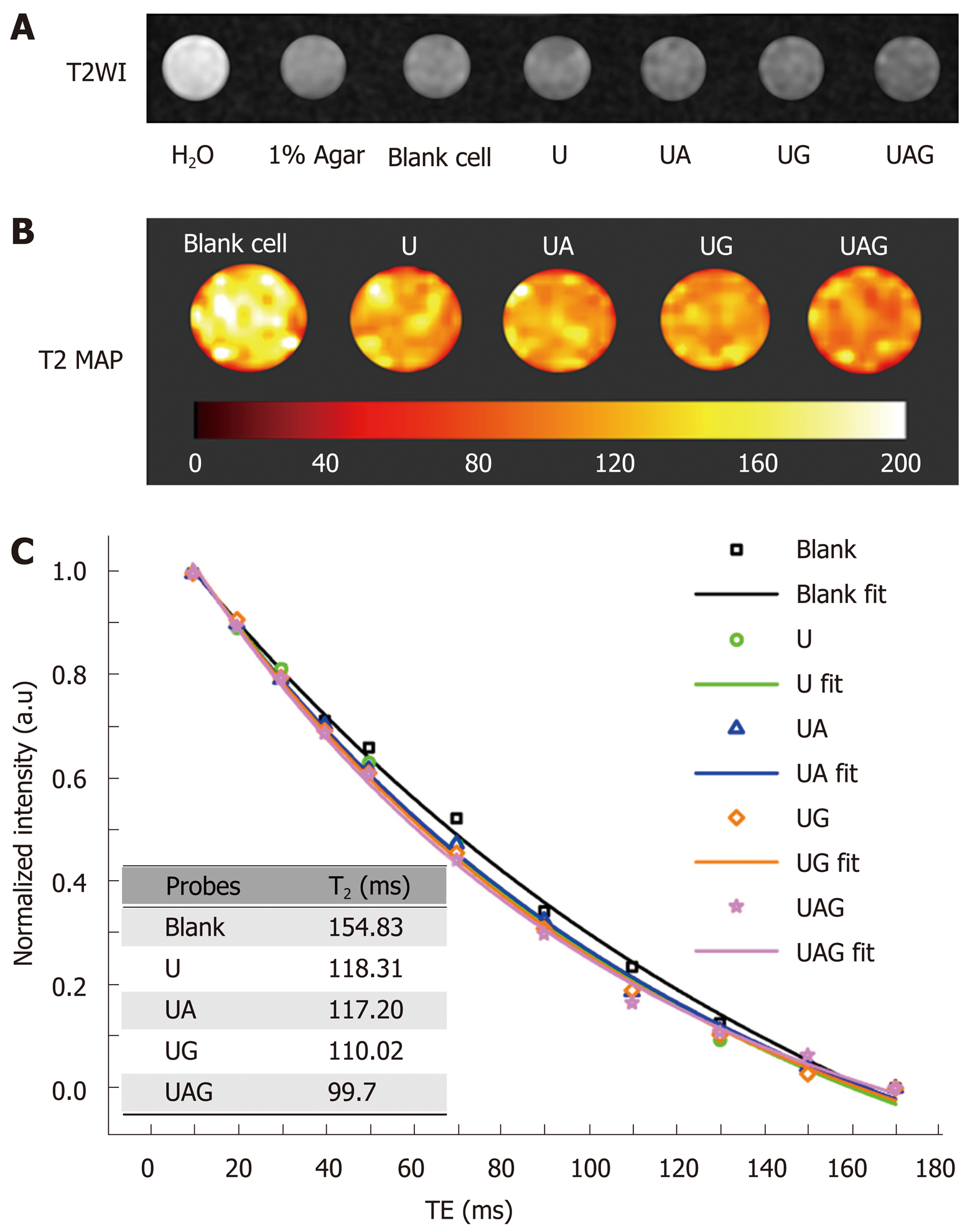
Figure 7 In vitro magnetic resonance imaging results demonstrating the binding efficiency and imaging properties of different ultra-small superparamagnetic iron oxide probes.
A: T2WI of different samples contained in a 96-well plate. From left to right: H2O, 1% agar, blank Hepa1-6/GPC3 cells, and hepa1-6/GPC3 cells treated with 100 µg Fe/mL USPIO (U), anti-AFP–USPIO (UA), anti-GPC3–USPIO (anti-GPC3–USPIO), or anti-AFP–USPIO–anti-GPC3 (UAG). B: Pseudocolor T2 map of cell samples treated with U, UA, UG, and UAG, respectively, compared with blank cells. T2 values are illustrated with a color bar. C: Signal intensities of cells after different probe treatments under different TE and exponential fits for the T2 values. Inset: The fitted T2 relaxation time for cells treated with different USPIO probes (blank control, U, UA, UG, or UAG). USPIO: Ultra-small superparamagnetic iron oxide; AFP: Alpha-fetoprotein; GPC3: Glypican-3; UAG: Anti-AFP–USPIO–anti-GPC3; UA: Anti-AFP–USPIO; UG: Anti-GPC3–USPIO; U: Unlabeled (non-targeted) USPIO; T2WI: T2-weighted imaging.
- Citation: Ma XH, Wang S, Liu SY, Chen K, Wu ZY, Li DF, Mi YT, Hu LB, Chen ZW, Zhao XM. Development and in vitro study of a bi-specific magnetic resonance imaging molecular probe for hepatocellular carcinoma. World J Gastroenterol 2019; 25(24): 3030-3043
- URL: https://www.wjgnet.com/1007-9327/full/v25/i24/3030.htm
- DOI: https://dx.doi.org/10.3748/wjg.v25.i24.3030


