Copyright
©The Author(s) 2019.
World J Gastroenterol. Jun 28, 2019; 25(24): 3030-3043
Published online Jun 28, 2019. doi: 10.3748/wjg.v25.i24.3030
Published online Jun 28, 2019. doi: 10.3748/wjg.v25.i24.3030
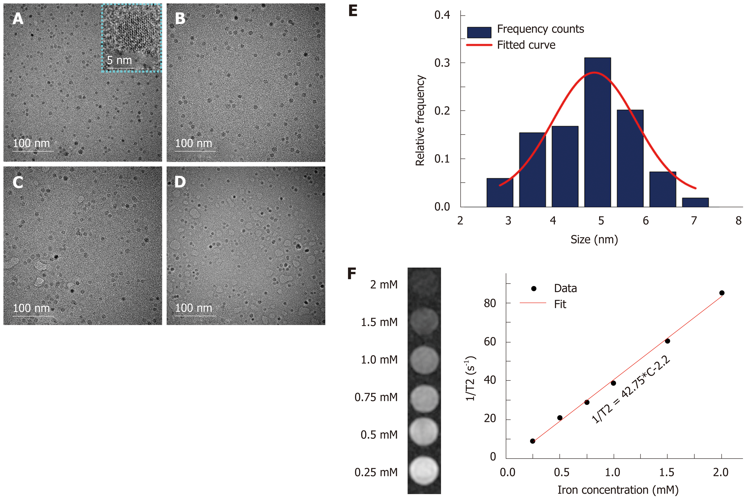
Figure 2 Physical characterization of ultra-small superparamagnetic iron oxide probes by transmission electron microscopy and magnetic resonance imaging.
A-D: Transmission electron microscopy (TEM) images of ultra-small superparamagnetic iron oxide (USPIO) probes of U, UA, UG, and UAG, respectively. E: The core size distribution of USPIO with a mean diameter of 4.88 nm and a standard deviation of 0.16 nm (n = 147), as determined from the TEM images. F: T2-weighted magnetic resonance images of a series of water solutions containing different concentrations of USPIO as indicated by iron concentration (left) and linear regression fitting of the transversal relaxation rate (1/T2) data vs different iron concentrations for extracting the transverse relaxivity r2 (right). UAG: Anti-AFP–USPIO–anti-GPC3; UA: Anti-AFP–USPIO; UG: Anti-GPC3–USPIO; U: Unlabeled (non-targeted) USPIO.
- Citation: Ma XH, Wang S, Liu SY, Chen K, Wu ZY, Li DF, Mi YT, Hu LB, Chen ZW, Zhao XM. Development and in vitro study of a bi-specific magnetic resonance imaging molecular probe for hepatocellular carcinoma. World J Gastroenterol 2019; 25(24): 3030-3043
- URL: https://www.wjgnet.com/1007-9327/full/v25/i24/3030.htm
- DOI: https://dx.doi.org/10.3748/wjg.v25.i24.3030


