Copyright
©The Author(s) 2019.
World J Gastroenterol. Jun 28, 2019; 25(24): 3030-3043
Published online Jun 28, 2019. doi: 10.3748/wjg.v25.i24.3030
Published online Jun 28, 2019. doi: 10.3748/wjg.v25.i24.3030
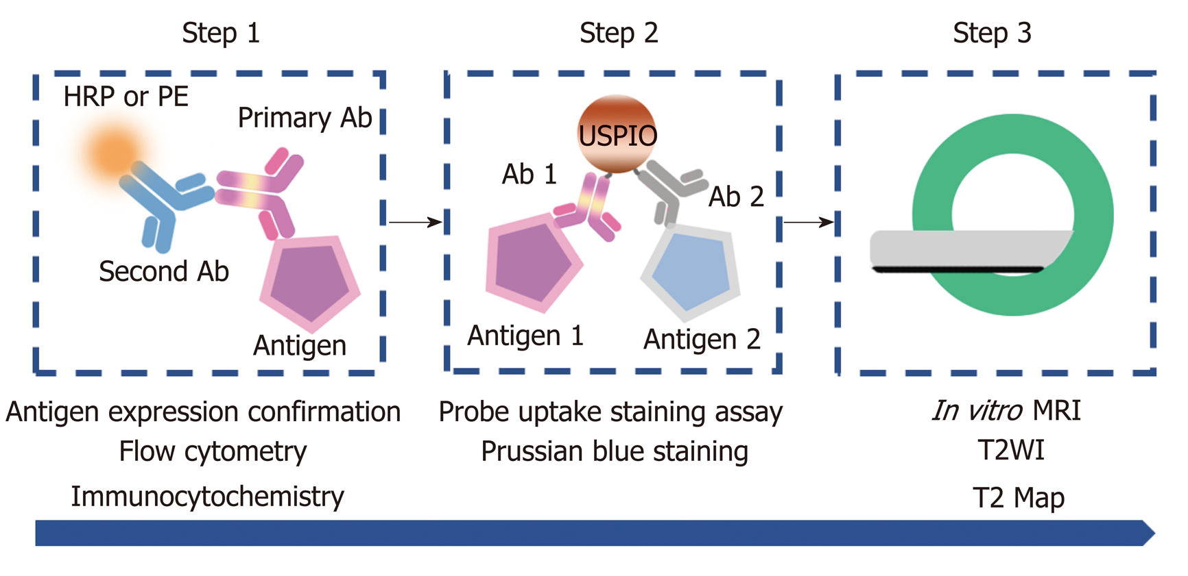
Figure 1 Flow of cell-based experiments.
First, AFP and GPC3 antigen expression on Hepa1-6/GPC3 cells was confirmed by flow cytometry and immunocytochemistry (step 1). Next, the cellular uptake of USPIO probes was investigated by performing Prussian blue-staining assays for iron (step 2). Finally, in vitro MRI was performed, including T2-weighted imaging (T2WI) and T2 Map imaging (step 3). HRP: Horseradish peroxidase; PE: Phycoerythrin; Ab: Antibody; USPIO: Ultra-small superparamagnetic iron oxide; MRI: Magnetic resonance imaging; T2WI: T2-weighted imaging.
- Citation: Ma XH, Wang S, Liu SY, Chen K, Wu ZY, Li DF, Mi YT, Hu LB, Chen ZW, Zhao XM. Development and in vitro study of a bi-specific magnetic resonance imaging molecular probe for hepatocellular carcinoma. World J Gastroenterol 2019; 25(24): 3030-3043
- URL: https://www.wjgnet.com/1007-9327/full/v25/i24/3030.htm
- DOI: https://dx.doi.org/10.3748/wjg.v25.i24.3030


