Copyright
©The Author(s) 2019.
World J Gastroenterol. Jun 21, 2019; 25(23): 2911-2923
Published online Jun 21, 2019. doi: 10.3748/wjg.v25.i23.2911
Published online Jun 21, 2019. doi: 10.3748/wjg.v25.i23.2911
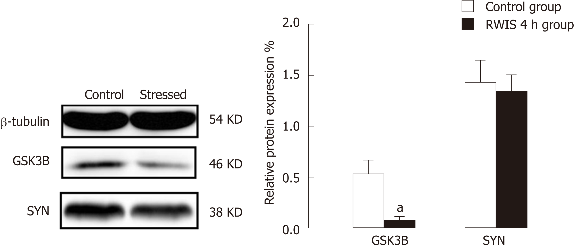
Figure 5 Verification of glycogen synthase kinase-3 beta and synaptophysin expression by Western blot.
After brain dissection, the proteins in the mediodorsal thalamic nucleu were separated by SDS-PAGE, and the expression levels of glycogen synthase kinase-3 beta (GSK3B) and synaptophysin (SYN) were detected by Western blot using antibodies against GSK3B and SYN. The bars represent the changes in the total GSK3B and SYN levels. Each value is the mean ± SD from at least three independent experiments; aP < 0.05. The expression of GSK3B was increased in the stressed group, whereas SYN showed no significant difference, which is in agreement with the isobaric tags for relative and absolute quantitation results.
- Citation: Gong SN, Zhu JP, Ma YJ, Zhao DQ. Proteomics of the mediodorsal thalamic nucleus of rats with stress-induced gastric ulcer. World J Gastroenterol 2019; 25(23): 2911-2923
- URL: https://www.wjgnet.com/1007-9327/full/v25/i23/2911.htm
- DOI: https://dx.doi.org/10.3748/wjg.v25.i23.2911


