Copyright
©The Author(s) 2019.
World J Gastroenterol. Jun 7, 2019; 25(21): 2636-2649
Published online Jun 7, 2019. doi: 10.3748/wjg.v25.i21.2636
Published online Jun 7, 2019. doi: 10.3748/wjg.v25.i21.2636
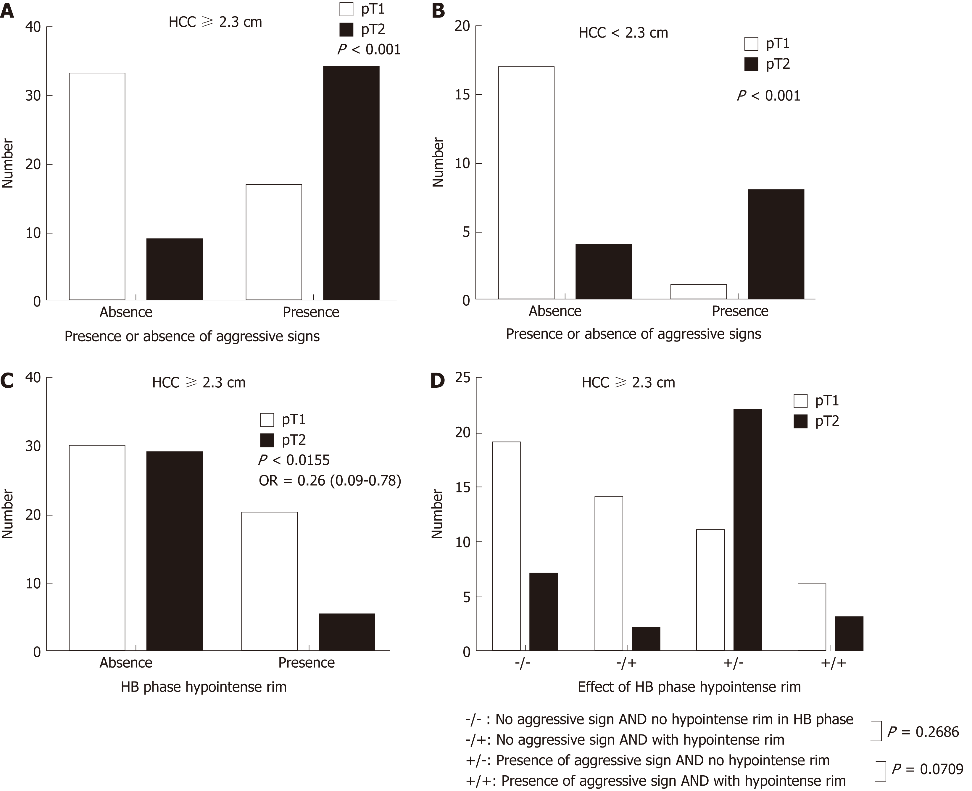
Figure 6 Solitary hepatocellular carcinomas on gadoxetic acid-enhanced magnetic resonance imaging.
Large (≥ 2.3 cm) (A) and small (< 2.3 cm) (B) solitary hepatocellular carcinomas (HCCs) with aggressive imaging findings tend to be stage pT2 tumors (P < 0.001). Large (≥ 2.3 cm) HCCs with hypointense rims on MRI in the HB phase tend to be a stage pT1 tumors (P = 0.0155); C: Large HCCs with aggressive imaging features are more likely to be stage pT2 tumors if a hypointense rim is absent rather than present in the hepatobiliary phase; D: HCC: Hepatocellular carcinoma; HB: Hepatobiliary; pT1: Pathologic stage T1; pT2: Pathologic stage T2.
- Citation: Chou YC, Lao IH, Hsieh PL, Su YY, Mak CW, Sun DP, Sheu MJ, Kuo HT, Chen TJ, Ho CH, Kuo YT. Gadoxetic acid-enhanced magnetic resonance imaging can predict the pathologic stage of solitary hepatocellular carcinoma. World J Gastroenterol 2019; 25(21): 2636-2649
- URL: https://www.wjgnet.com/1007-9327/full/v25/i21/2636.htm
- DOI: https://dx.doi.org/10.3748/wjg.v25.i21.2636


