Copyright
©The Author(s) 2019.
World J Gastroenterol. Jun 7, 2019; 25(21): 2623-2635
Published online Jun 7, 2019. doi: 10.3748/wjg.v25.i21.2623
Published online Jun 7, 2019. doi: 10.3748/wjg.v25.i21.2623
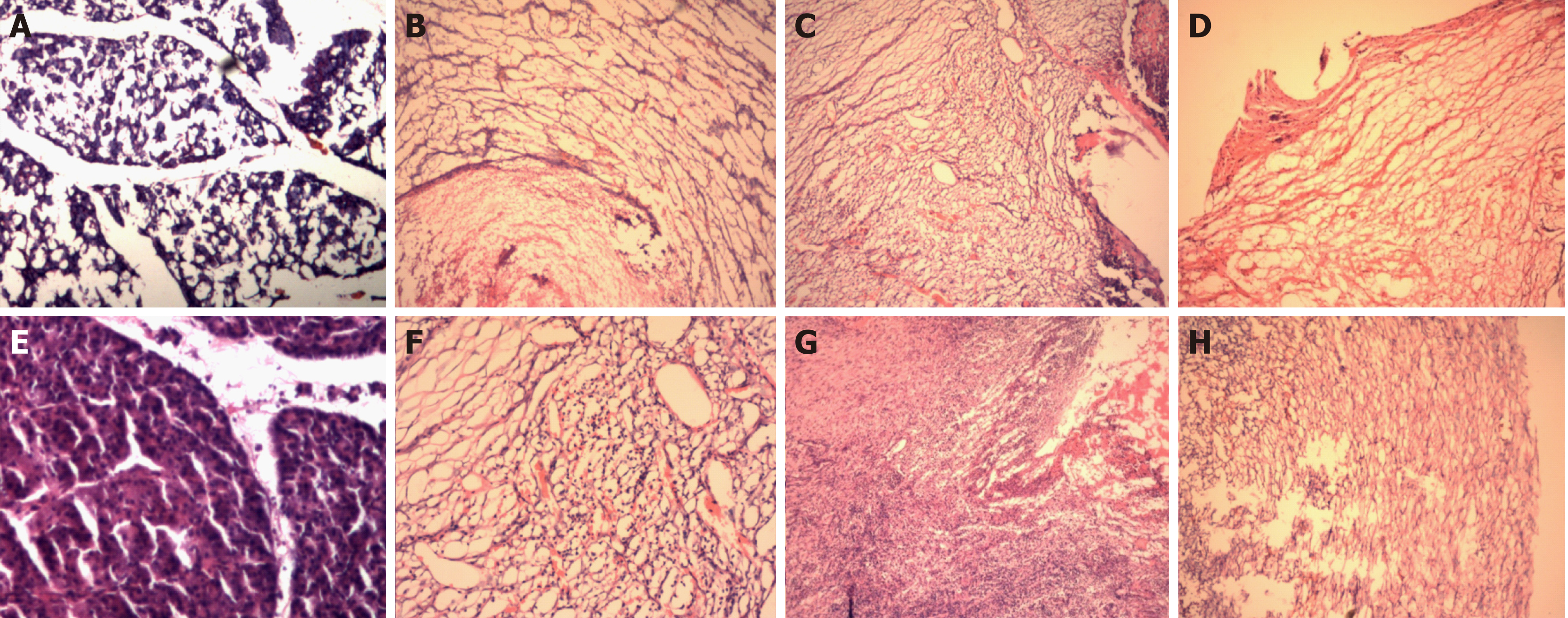
Figure 8 Hematoxylin and eosin staining.
A: Hematoxylin and eosin (HE) staining of the normal pancreas in the control group (40×); B: HE stained image (40×) on the 3rd day postoperatively in experimental group one; C: HE stained image (40×) on the 7th day postoperatively in experimental group one. D: HE stained image (40×) on the 14th day postoperatively in experimental group one. E: HE staining of the normal pancreas in the control group (40×); F: HE stained image (40×) on the 3rd day postoperatively in experimental group two; G: HE stained image (40×) on the 7th day postoperatively in experimental group two. H: HE stained image (40×) on the 14th day postoperatively in experimental group two.
- Citation: Wang S, Zhang K, Hu JL, Wu WC, Liu X, Ge N, Guo JT, Wang GX, Sun SY. Endoscopic resection of the pancreatic tail and subsequent wound healing mechanisms in a porcine model. World J Gastroenterol 2019; 25(21): 2623-2635
- URL: https://www.wjgnet.com/1007-9327/full/v25/i21/2623.htm
- DOI: https://dx.doi.org/10.3748/wjg.v25.i21.2623


