Copyright
©The Author(s) 2019.
World J Gastroenterol. Jun 7, 2019; 25(21): 2623-2635
Published online Jun 7, 2019. doi: 10.3748/wjg.v25.i21.2623
Published online Jun 7, 2019. doi: 10.3748/wjg.v25.i21.2623
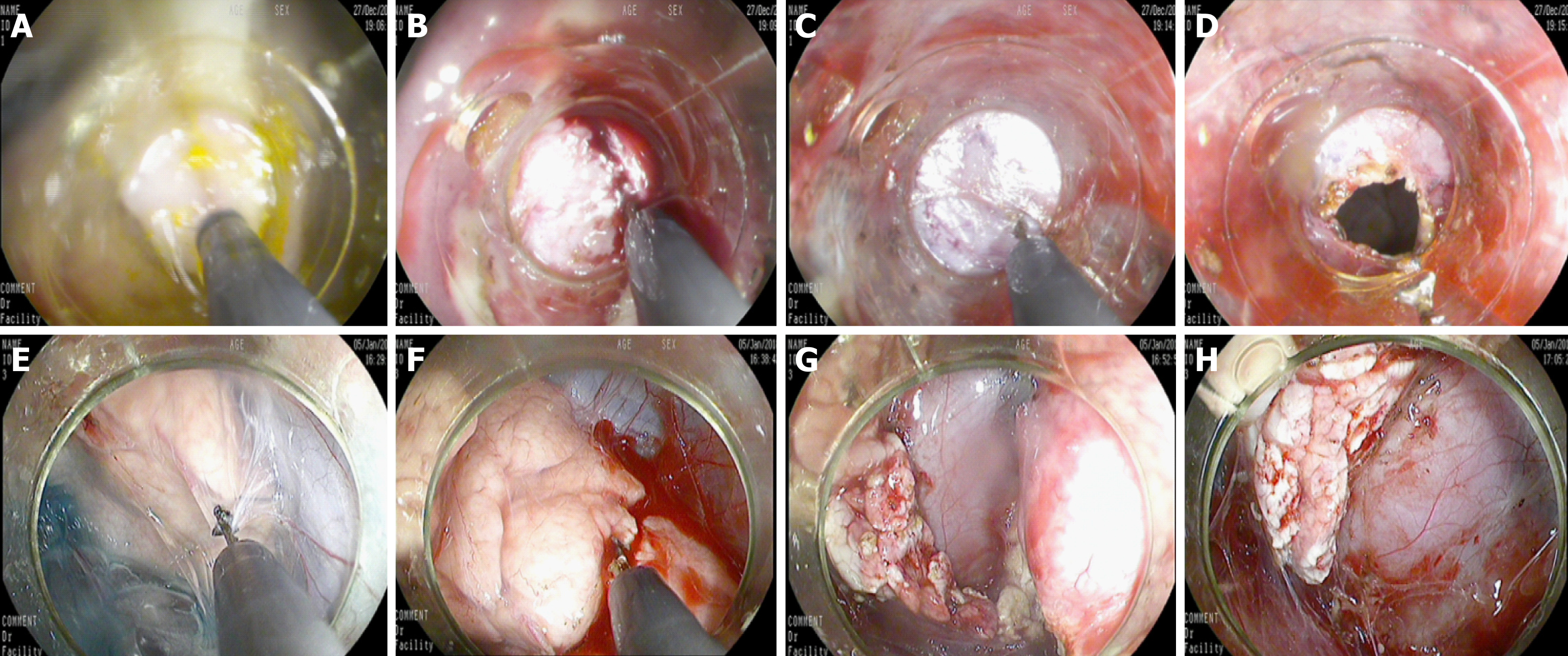
Figure 2 The mucosal, submucosal, and muscular layers were sequentially cut with a triangular knife until the retroperitoneum was reached.
A: The incising of the mucosa of the gastric wall; B: The incising of the submucosa of the gastric wall; C: The incising of the muscular layer of the gastric wall; D: The incising of the serosal layer of the gastric wall; E: The separation of the pancreatic capsule; F: The incision of the parenchyma of the pancreatic body and tail; G: Cutting the pancreatic tail; H: The residual pancreatic body wound after removal of the pancreatic tail.
- Citation: Wang S, Zhang K, Hu JL, Wu WC, Liu X, Ge N, Guo JT, Wang GX, Sun SY. Endoscopic resection of the pancreatic tail and subsequent wound healing mechanisms in a porcine model. World J Gastroenterol 2019; 25(21): 2623-2635
- URL: https://www.wjgnet.com/1007-9327/full/v25/i21/2623.htm
- DOI: https://dx.doi.org/10.3748/wjg.v25.i21.2623


